


Validation of an in vitro biological method for filgrastim potency assessment

https://doi.org/10.30895/2221-996X-2026-26-1-85-96

Abstract
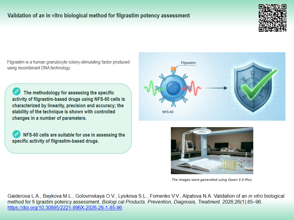
INTRODUCTION. Filgrastim is a human granulocyte colony-stimulating factor produced using recombinant DNA technology. Since filgrastim quality control necessitates a validated method, establishing the entire method validation parameters is of high priority; confirming the previously established parameters gains primary importance when substituting critical reagents/ materials/parameters.
AIM. This study aimed to select and confirm a number of validation parameters for in vitro biological method that would assess filgrastim potency using NFS-60 cell line.
MATERIALS AND METHODS. Filgrastim potency was assessed by fluorescence intensity of alamarBlue™ reagent at excitation wavelengths of 530 nm and emission of 620 nm directly proportional to proliferation level of NFS-60 cells exposed to filgrastim 0.1 to 208 IU/mL. Statistical analysis of the results was performed using the 4-parameter logistic function 4PL and the parallel line analysis in PLA 2.0.0 software.
RESULTS. The method was deemed precise: the coefficient of variation (CV) in the repeatability study was 6.20%, and the intermediate precision study showed CV 1.19% (acceptance criterion CV≤25%). The method was linear; the coefficient of determination for the linear function was R2=0.99 (acceptance criterion R2≥0.95). The degree of recovery was 101.6% and did not exceed ±10% of the expected value. The method was robust to an increased density of the cell suspension from 1.5×10⁵ to 3.0×10⁵ cells/mL (CV=0.07%) and to prolonged incubation of filgrastim samples with the cell suspension (from 48 to 72 h, CV=4.2%) or with fluorescent dye (from 4 to 6 h, CV=2.05%).
CONCLUSIONS. The in vitro method assessing specific potency of filgrastim-based preparations using NFS-60 cell line is linear, precise, and accurate, and has proven to be stable under controlled changes of certain parameters. Confirmed applicability of NFS-60 cell line for assessing the potency of filgrastim-based preparations is essential for manufacturers, since it expands the range of cell cultures used for quality assessment of preparations.
Keywords
specific activity,
biological method,
validation parameters,
correctness,
linearity,
robustness,
precision,
NFS-60 cell line,
granulocyte colony-stimulating factor
Funding: The study was conducted by the Scientific Centre for Expert Evaluation of Medicinal Products as part of the applied research funded under State Assignment No. 056-00061-26-00 (R&D Registry No. 124022200103-5)
About the Authors
L. A. Gaiderova
Scientific Centre for Expert Evaluation of Medicinal Products
Russian Federation
Russian Federation
Lidia A. Gaiderova, Cand. Sci. (Med.)
8/2 Petrovsky Blvd, Moscow 127051
M. L. Baykova
Scientific Centre for Expert Evaluation of Medicinal Products
Russian Federation
Russian Federation
Marina L. Baykova
8/2 Petrovsky Blvd, Moscow 127051
O. V. Golovinskaya
Scientific Centre for Expert Evaluation of Medicinal Products
Russian Federation
Russian Federation
Olga V. Golovinskaya, Cand. Sci. (Med.)
8/2 Petrovsky Blvd, Moscow 127051
S. L. Lysikova
Scientific Centre for Expert Evaluation of Medicinal Products
Russian Federation
Russian Federation
Svetlana L. Lysikova, Cand. Sci. (Med.)
8/2 Petrovsky Blvd, Moscow 127051
V. V. Fomenko
Scientific Centre for Expert Evaluation of Medicinal Products
Russian Federation
Russian Federation
Viktoriia V. Fomenko
8/2 Petrovsky Blvd, Moscow 127051
N. A. Alpatova
Scientific Centre for Expert Evaluation of Medicinal Products
Russian Federation
Russian Federation
Natalia А. Alpatova, Dr. Sci. (Biol.)
8/2 Petrovsky Blvd, Moscow 127051
References
1. Trinh NTM, Thuoc TL, Thao DTP. Production of recombinant human G-CSF from non-classical inclusion bodies in Escherichia coli. Braz J Microbiol. 2021;52(2):541–6. https://doi.org/10.1007/s42770-020-00413-y
2. Bond TC, Szabo E, Gabriel S, et al. Meta-analysis and indirect treatment comparison of lipegfilgrastim with pegfilgrastim and filgrastim for the reduction of chemotherapyinduced neutropenia-related events. J Oncol Pharm Pract. 2018;24(6):412–23. https://doi.org/10.1177/1078155217714859
3. Cornes P, Gascon P, Chan St, et al. Systematic review and meta-analysis of short-versus long-acting granulocyte colony-stimulating factors for reduction of chemotherapy-induced febrile neutropenia. Adv Ther. 2018;35(11):1816–29. https://doi.org/10.1007/s12325-018-0798-6
4. Levy MJ, Gucinski AC, Sommers CD, et al. Analytical techniques and bioactivity assays to compare the structure and function of filgrastim (granulocyte-colony stimulating factor) therapeutics from different manufacturers. Anal Bioanal Chem. 2014;406(26):6559–67. https://doi.org/10.1007/s00216-013-7469-x
5. Magalhaes V, Mantovani M, Caruso C, et al. Physicochemical and biological comparison of the first Brazilian biosimilar filgrastim with its reference product. Biosimilars. 2016;6:45–60. https://doi.org/10.2147/BS.S107898
6. Caselli D, Cesaro S, Aricò M. Biosimilars in the management of neutropenia: Focus on filgrastim. Biologics. 2016;10:17–22. https://doi.org/10.2147/BTT.S73580
7. Avdeeva ZhI, Soldatov AA, Alpatova NA, et al. Recombinant granulocyte colony stimulating factor biosimilars. Quality assessment. BIOpreparation. Prevention, Diagnosis, Treatment. 2015;(1):4–14 (In Russ.). EDN: UBEKFJ
8. White JR, Abodeely M, Ahmed S, et al. Best practices in bioassay development to support registration of biopharmaceuticals. Biotechniques. 2019;67(3):126–37. https://doi.org/10.2144/btn-2019-0031
9. Li H, Witkos TM, Umlauf S, Thompson C. Potency assay variability estimation in practice. Pharm Stat. 2025;24(1):e2408. https://doi.org/10.1002/pst.2408
10. Präbst K, Engelhardt H, Ringgeler S, Hübner H. Basic сolorimetric proliferation assays: MTT, WST, and Resazurin. Methods Mol Biol. 2017;1601:1–17. https://doi.org/10.1007/978-1-4939-6960-9_1
11. Kumar P, Nagarajan A, Uchil PD. Analysis of cell viability by the MTT assay. Cold Spring Harb Protoc. 2018;2018(6):10. https://doi.org/10.1101/pdb.prot095505
12. Tiwari K, Wavdhane M, Haque S, et al. A sensitive WST8-based bioassay for PEGylated granulocyte colony stimulating factor using the NFS-60 cell line. Pharm Biol. 2015;53(6):849–54. https://doi.org/10.3109/13880209.2014.943248
13. Kumar P, Nagarajan A, Uchil PD. Analysis of cell viability by the alamarBlue assay. Cold Spring Harb Protoc. 2018;2018(6):10. https://doi.org/10.1101/pdb.prot095489
14. Golovinskaya OV, Baykova ML, Alpatova NA, et al. Comparative analysis of dyes used in the assessment of filgrastim products specific activity by biological in vitro methods. BIOpreparations. Prevention, Diagnosis, Treatment. 2020;20(3):193–201 (In Russ.). https://doi.org/10.30895/2221-996X-2020-20-3-193-201
15. Nakoinz I, Lee MT, Weaver JF, Ralph P. Differentiation of the IL-3-dependent NFS-60 cell line and adaption to growth in macrophage colony-stimulating factor. J Immunol. 1990;145(3):860–4. PMID: 2142710
16. Wadhwa M, Bird C, Hamill M, et al. The 2nd International Standard for human granulocyte colony stimulating factor. J Immunol Methods. 2011;367(1–2):63–9. https://doi.org/10.1016/j.jim.2011.02.005
17. Nikonova YuA, Abbasova SG, Kargopolova PE, et al. Validation of an analytical procedure for evaluating the biological activity of a medicinal product based on tocilizumab and determination of acceptance criteria for test results. Regulatory Research and Medicine Evaluation. 2024;14(3):317–29 (In Russ.). https://doi.org/10.30895/1991-2919-2024-14-3-317-329
18. Kirkland PD, Newberry KM. Your assay has changed — is it still “fit for purpose”? What evaluation is required. Rev Sci Tech. 2021;40(1):205–15. https://doi.org/10.20506/rst.40.1.3218
Supplementary files

|
1. Fig. S1. Example of a dose–response curve for filgrastim concentration (potency) range from 0.52 to 0.0005 ng/mL (0.05–52 IU/mL). NFS-60 cell line was used in the study. X-axis, common logarithm of filgrastim doses; Y-axis, fluorescence signals. A, mean graph; В, unrestricted model; С, restricted model. RFU, relative fluorescence units. | |
| Subject | ||
| Type | Исследовательские инструменты | |
Download
(385KB)
|
Indexing metadata ▾ | |
|
|
2. Fig. S2. An example of a linear part of the dose–response curve evaluating filgrastim potency and using NFS-60 cell line. X-axis, common logarithm of filgrastim doses; Y-axis, fluorescence signals. RFU, relative fluorescence units. | |
| Subject | ||
| Type | Исследовательские инструменты | |
Download
(194KB)
|
Indexing metadata ▾ | |
|
|
3. Fig. S3. Graph of the expected filgrastim relative potency versus the values obtained in the study. The graph demonstrates the method linearity. R², coefficient of determination. | |
| Subject | ||
| Type | Исследовательские инструменты | |
Download
(120KB)
|
Indexing metadata ▾ | |
|
|
4. Table S1. Characterization of a linear part of fluorescence intensity versus filgrastim doses graph | |
| Subject | ||
| Type | Исследовательские инструменты | |
Download
(114KB)
|
Indexing metadata ▾ | |
Review
For citations:
Gaiderova L.A., Baykova M.L., Golovinskaya O.V., Lysikova S.L., Fomenko V.V., Alpatova N.A. Validation of an in vitro biological method for filgrastim potency assessment. Biological Products. Prevention, Diagnosis, Treatment. 2026;26(1):85-96. (In Russ.) https://doi.org/10.30895/2221-996X-2026-26-1-85-96
Views:
420
JATS XML





 Email this article