


Long-term stability of liposomal mycobacteriophage D29: A comprehensive in vitro assessment

https://doi.org/10.30895/2221-996X-2026-26-1-67-74

Abstract
INTRODUCTION. The spread of antibiotic-resistant mycobacteria necessitates new therapeutic approaches for tuberculosis, phage therapy being one of them. Liposomal mycobacteriophages are considered to be a way to enhance their efficacy; however, a comprehensive assessment of their stability is warranted for the further use of such preparations.
AIM. This study aimed to evaluate the long-term stability (over six months) of a liposomal form of mycobacteriophage D29 under storage conditions at 4 °C based on a set of physicochemical and functional parameters to substantiate its further preclinical study.
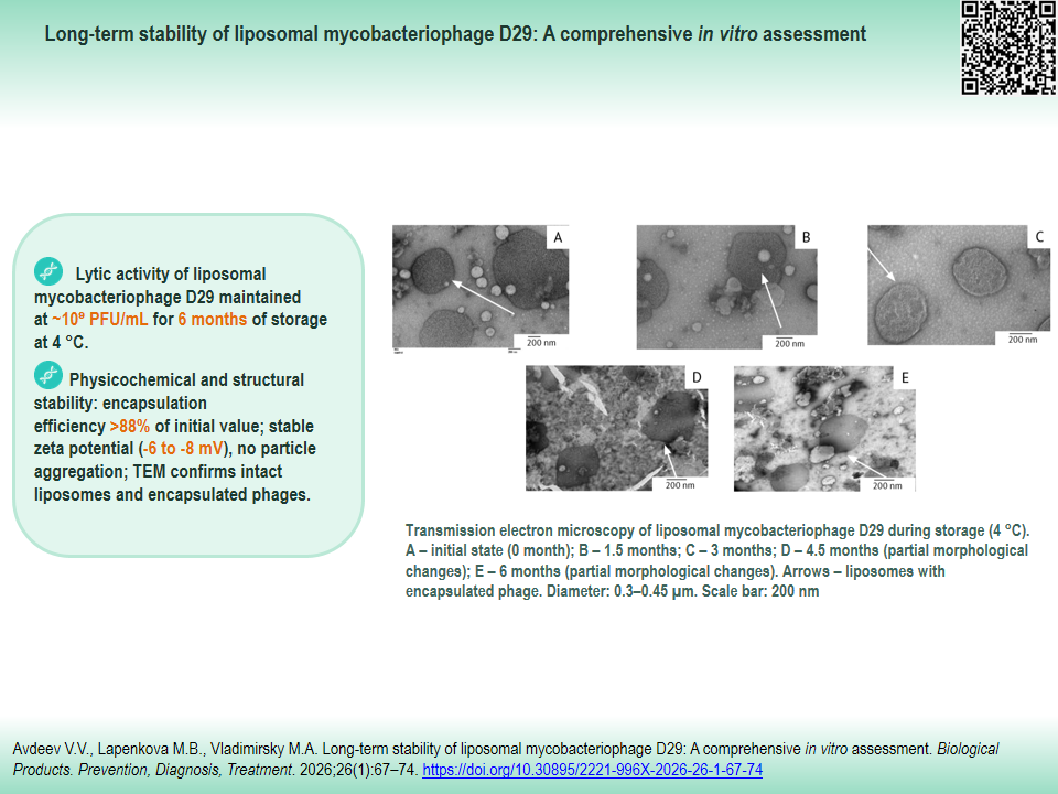
MATERIALS AND METHODS. The stability of liposomal form D29 was assessed over six months of storage at 4 °C. Sampling was performed at the following time points: 0; 1.5; 3; 4.5; and 6 months. The degree of phage encapsulation (%) was quantified by real-time polymerase chain reaction (qPCR). The surface charge (zeta potential) was measured by electrophoretic light scattering (Doppler electrophoretic mobility). Liposome morphology was analyzed using transmission electron microscopy. Functional activity was assessed by determining lytic activity via the Grazia titration method on M. smegmatis mc² 155 culture.
RESULTS. High stability of the liposomal phage D29 form was demonstrated. Lytic activity remained at a level of ~109 PFU/mL throughout the storage period (six months, 4 °C). The degree of encapsulation decreased insignificantly (>88% of the initial value). The constant zeta potential value (from –6.1±0.3 to –8.2±0.5 mV) confirmed colloidal stability of the suspension without particle aggregation throughout the entire storage period.
CONCLUSIONS. The liposomal mycobacteriophage D29 demonstrates high physicochemical and functional stability after long-term storage (six months, 4 °C), with preserved lytic activity and a high degree of encapsulation. The findings substantiate the prospects for further preclinical and subsequent clinical trial of this preparation designed to treat mycobacterial infections.
Keywords
mycobacteriophage D29,
liposomes,
drug stability,
encapsulation,
zeta potential,
transmission electron microscopy,
long-term storage,
lytic activity
Funding: The work was carried out as part of the state assignment of the National Medical Research Center of Phthisiopulmonology and Infectious Diseases of the Ministry of Health of the Russian Federation, registration No. NIOTKR 121031600067-1 Development of a New Anti-Tuberculosis Drug Based on the Liposomal Mycobacteriophage D29 for the Treatment of Pulmonary Tuberculosis, Including Drug-Resistant Forms
About the Authors
V. V. Avdeev
National Medical Research Center of Phthisiopulmonology and Infectious Diseases of the Ministry of Health of the Russian Federation
Russian Federation
Russian Federation
Vadim V. Avdeev
4/1 Dostoevsky St., Moscow 127473
M. B. Lapenkova
National Medical Research Center of Phthisiopulmonology and Infectious Diseases of the Ministry of Health of the Russian Federation
Russian Federation
Russian Federation
Marina B. Lapenkova, Cand. Sci. (Med.)
4/1 Dostoevsky St., Moscow 127473
M. A. Vladimirsky
National Medical Research Center of Phthisiopulmonology and Infectious Diseases of the Ministry of Health of the Russian Federation
Russian Federation
Russian Federation
Mikhail A. Vladimirsky, Dr. Sci. (Med.), Prof.
4/1 Dostoevsky St., Moscow 127473
References
1. Dean AS, Tosas Auguet O, Glaziou P, et al. 25 years of surveillance of drug-resistant tuberculosis: achievements, challenges, and way forward. Lancet Infect Dis. 2022;22(7):e191–6. https://doi.org/10.1016/s1473-3099(21)00808-2
2. Sterlikov SA, Vasilyeva IA, Mikhaylova YuV, et al. The new definition and epidemiology of extensive drug resistant tuberculosis in 2020. Tuberculosis and Lung Diseases. 2023;101(2):14–9 (In Russ.). https://doi.org/10.58838/2075-1230-2023-101-2-14-19
3. Vasilyeva IA, Panova AE, Tinkova VV, et al. Antimicrobial resistance of Mycobacterium avium during the COVID-19 pandemic. Clinical Microbiology and Antimicrobial Chemotherapy. 2024;26(4):462–9. https://doi.org/10.36488/cmac.2024.4.462-469
4. Mozhokina GN, Zyuzya YuR, Petrova LYu, et al. Toxicity of treatment regimens for drugresistant tuberculosis. Antibiotics and Chemotherapy. 2021;66(11–12):25–30 (In Russ.). https://doi.org/10.37489/0235-2990-2021-66-11-12-25-30
5. Abramchenko AV, Romanova MI, Gayda AI, et al. Effectiveness and safety of short-course chemotherapy regimens for drug-resistant tuberculosis: Literature review and meta-analysis. Tuberculosis and Lung Diseases. 2025;103(2):26–37 (In Russ.). https://doi.org/10.58838/2075-1230-2025-103-2-26-37
6. Vasilyeva IA. Achievements and prospects of innovative research in the field of phthisiology. Herald of the Russian Academy of Sciences. 2025;(1):63–74 (In Russ.). https://doi.org/10.7868/S3034520025010063
7. Abedon ST, Kuhl SJ, Blasdel BG, Kutter EM. Phage treatment of human infections. Bacteriophage. 2011;1(2):66–85. https://doi.org/10.4161/bact.1.2.15845
8. Lin DM, Koskella B, Lin HC. Phage therapy: An alternative to antibiotics in the age of multi-drug resistance. World J Gastrointest Pharmacol Ther. 2017;8(3):162–73. https://doi.org/10.4292/wjgpt.v8.i3.162
9. Jacobs-Sera D, Hatfull GF. On the nature of mycobacteriophage diversity and host preference. Virology. 2012;434(2):187–201 https://doi.org/10.1016/j.virol.2012.09.026
10. Pope WH, Bowman CA, Russell DA, et al. Expanding the diversity of mycobacteriophages: insights into genome architecture and evolution. PLoS One. 2011;6(1):e16329. https://doi.org/10.1371/journal.pone.0016329
11. Flint R, Laucirica DR, Chan HK, et al. Stability considerations for bacteriophages in liquid formulations designed for nebulization. Cells. 2023;12(16):2057. https://doi.org/10.3390/cells12162057
12. Allen TM, Cullis PR. Liposomal drug delivery systems: from concept to clinical applications. Adv Drug Deliv Rev. 2013;65(1):36–48. https://doi.org/10.1016/j.addr.2012.09.037
13. Colom J, Cano-Sarabia M, Otero J, et al. Liposome-encapsulated bacteriophages for enhanced oral phage therapy against Salmonella spp. Appl Environ Microbiol. 2015;81(14):4841–9. https://doi.org/10.1128/AEM.00812-15
14. Kumar A, Arun JK, Matta Y, et al. Optimization of nanoliposome formulations for targeted delivery of hydrophobic drugs. Front Health Inform. 2024;13(7):745–57.
15. Sawant RR, Torchilin VP. Challenges in development of targeted liposomal therapeutics. AAPS J. 2012;14(2):303–15. https://doi.org/10.1208/s12248-012-9330-0
16. Leung SSY, Parumasivam T, Gao FG, et al. Production of inhalation phage powders using spray freeze drying and spray drying techniques for treatment of respiratory infections. Pharm Res. 2016;33(6):1486–96. https://doi.org/10.1007/s11095-016-1892-6
17. Avdeev VV, Kuzin VV, Vladimirsky MA, Vasilieva IA. Experimental studies of the liposomal form of lytic mycobacteriophage D29 for the treatment of tuberculosis infection. Microorganisms. 2023;11(5):1214. https://doi.org/10.3390/microorganisms11051214
18. Avdeev VV, Vladimirsky MA, Samoilova AG, Vasilieva IA. Therapeutic efficacy against a drug-resistant strain of M. tuberculosis in an experimental study on inbred mice and associated toxicity evaluation. Phage (New Rochelle). 2025;6(2). https://doi.org/10.1089/phage.2024.0066
19. Šimoliūnas E, Vilkaitytė M, Kaliniene L, et al. Incomplete LPS Core-Specific Felix01-Like Virus vB_EcoM_ VpaE1. Viruses. 2015;7(12):6163–81. https://doi.org/10.3390/v7122932
20. Cinquerrui S, Mancuso F, Vladisavljević GT, et al. Nanoencapsulation of bacteriophages in liposomes prepared using microfluidic hydrodynamic flow focusing. Front Microbiol. 2018;9:2172. https://doi.org/10.3389/fmicb.2018.02172
21. Biltonen RL, Lichtenberg D. The use of differential scanning calorimetry as a tool to characterize liposome preparations. Chem Phys Lipids. 1993;64(1–3):129–42. https://doi.org/10.1016/0009-3084(93)90062-8
22. Steven AC. Visualization of virus structure in three dimensions. Methods Cell Biol. 1981;22:297–323. https://doi.org/10.1016/S0091-679X(08)61881-6
23. Cullis PR, Hope MJ. Lipid nanoparticle systems for enabling gene therapies. Mol Ther. 2017;25(7):1467–75. https://doi.org/10.1016/j.ymthe.2017.03.013
24. Immordino ML, Dosio F, Cattel L. Stealth liposomes: review of the basic science, rationale, and clinical applications, existing and potential. Int J Nanomedicine. 2006;1(3):297–315. PMID: 17717971
Review
For citations:
Avdeev V.V., Lapenkova M.B., Vladimirsky M.A. Long-term stability of liposomal mycobacteriophage D29: A comprehensive in vitro assessment. Biological Products. Prevention, Diagnosis, Treatment. 2026;26(1):67-74. (In Russ.) https://doi.org/10.30895/2221-996X-2026-26-1-67-74
Views:
529
JATS XML





 Email this article