


Development of acid alpha-glucosidase-producing clones based on the CHO-K1 cell line
 S. S. Timonova,
S. S. Shubina,
J. S. Snegireva,
R. L. Anisimov,
N. V. Nikiforova,
D. A. Tretyak,
M. A. Koroleva,
M. Yu. Neronova,
R. A. Khamitov
S. S. Timonova,
S. S. Shubina,
J. S. Snegireva,
R. L. Anisimov,
N. V. Nikiforova,
D. A. Tretyak,
M. A. Koroleva,
M. Yu. Neronova,
R. A. Khamitov https://doi.org/10.30895/2221-996X-2026-26-2-171-182

Abstract
INTRODUCTION. The development of a biopharmaceutical drug for enzyme replacement therapy based on recombinant acid α-1,4-glucosidase (α-glucosidase) is a pressing task, the solution of which could provide patients with Pompe disease in the Russian Federation with the necessary amount of the drug. This study demonstrates an effective method for developing, from the CHO-K1 cell line, a stable industrial producer clone that produces active α-glucosidase.
AIM. This study aimed to develop monoclonal cell lines producing recombinant acid α-glucosidase and to evaluate the stability of growth characteristics and productivity during cultivation over 60 generations.
MATERIALS AND METHODS. The suspension CHO-K1 cell line (ECACC) was cultured in BalanCD Growth A medium. Cell transfection was performed by electroporation using the MaxCyte system (according to the CHO protocol). Selection of producers was carried out using puromycin (5 μg/mL). Cloning was performed using a cell dispensing system based on microfluidic technology (C.SIGHT). The monoclonality of the cell lines was confirmed using an automated cell imaging system (Cell Metric CLD). The concentration of α-glucosidase in the culture fluid was determined by enzyme-linked immunosorbent assay. Enzyme activity was measured by a colorimetric method using the substrate 4-nitrophenyl-α-D-glucopyranoside (pNP-α-D-Glc).
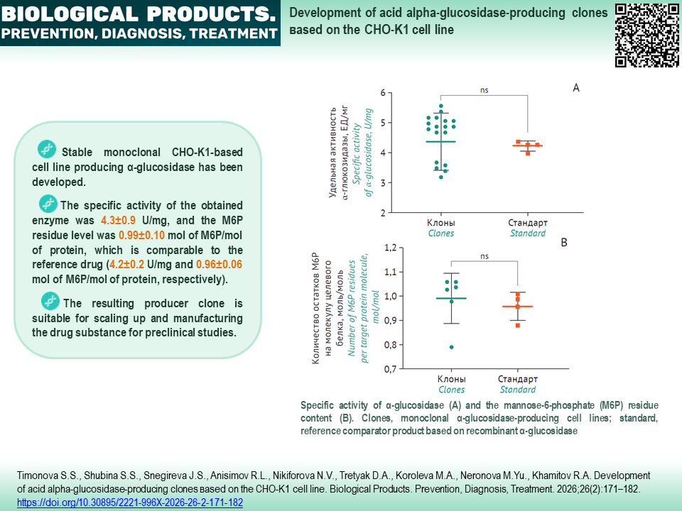
RESULTS. Screening of 1000 producers was performed, resulting in the selection of 22 cell lines with a productivity of 0.14–0.65 g/L. The lead α-glucosidase producer GAA-14 was cloned, followed by confirmation of monoclonality. A panel of 20 monoclonal lines was obtained. The specific activity of the enzyme and the mannose-6-phosphate residue (М6Р) content did not differ from those of the reference drug (4.3±0.9 U/mg and 0.99±0.10 mol M6P/mol protein, respectively). When studying the stability of the lead clone over 60 generations, preservation of growth characteristics (viability, 98.5±1.5%; population doubling time, 20.0±1.3 h; and productivity, 450±20 mg/L) was demonstrated under batch cultivation over 7 days.
CONCLUSIONS. A stable monoclonal α-glucosidase producer cell line based on CHO-K1 cells has been developed, yielding the active enzyme at 0.45 g/L on day 7. The resulting producer clone is suitable for scale-up and manufacture of the drug substance for preclinical studies.
Keywords
CHO-K1 cell line,
monoclonal cell line,
acid alpha-glucosidase,
lysosomal acid alpha-1,4-glucosidase,
4-nitrophenyl-α-D-glucopyranoside,
glycogen storage disease type II,
GSD II,
recombinant enzymes
Funding: The study was funded by GENERIUM JSC
About the Authors
S. S. Timonova
GENERIUM JSC
Russian Federation
Russian Federation
Sofia S. Timonova, Cand. Sci. (Biol.)
14 Vladimirskaya St., Volginsky, Pokrov, Vladimir Region 601125
S. S. Shubina
GENERIUM JSC
Russian Federation
Russian Federation
Sofia S. Shubina
14 Vladimirskaya St., Volginsky, Pokrov, Vladimir Region 601125
J. S. Snegireva
GENERIUM JSC
Russian Federation
Russian Federation
Julia S. Snegireva
14 Vladimirskaya St., Volginsky, Pokrov, Vladimir Region 601125
R. L. Anisimov
GENERIUM JSC
Russian Federation
Russian Federation
Roman L. Anisimov, Cand. Sci. (Biol.)
14 Vladimirskaya St., Volginsky, Pokrov, Vladimir Region 601125
N. V. Nikiforova
GENERIUM JSC
Russian Federation
Russian Federation
Natalya V. Nikiforova
14 Vladimirskaya St., Volginsky, Pokrov, Vladimir Region 601125
D. A. Tretyak
GENERIUM JSC
Russian Federation
Russian Federation
Danila A. Tretyak
14 Vladimirskaya St., Volginsky, Pokrov, Vladimir Region 601125
M. A. Koroleva
GENERIUM JSC
Mariya A. Koroleva
14 Vladimirskaya St., Volginsky, Pokrov, Vladimir Region 601125
M. Yu. Neronova
GENERIUM JSC
Russian Federation
Russian Federation
Maria Yu. Neronova
14 Vladimirskaya St., Volginsky, Pokrov, Vladimir Region 601125
R. A. Khamitov
GENERIUM JSC
Russian Federation
Russian Federation
Ravil A. Khamitov, Dr. Sci. (Med.), Prof.
14 Vladimirskaya St., Volginsky, Pokrov, Vladimir Region 601125
References
1. Herzog A, Hartung R, Reuser AJ, et al. A cross-sectional single-centre study on the spectrum of Pompe disease, German patients: molecular analysis of the GAA gene, manifestation and genotype-phenotype correlations. Orphanet J Rare Dis. 2012;7:35. https://doi.org/10.1186/1750-1172-7-35
2. Wan L, Lee CC, Hsu CM, et al. Identification of eight novel mutations of the acid alpha-glucosidase gene causing the infantile or juvenile form of glycogen storage disease type II. J Neurol. 2008;255(6):831–8. https://doi.org/10.1007/s00415-008-0714-0
3. Taverna S, Cammarata G, Colomba P, et al. Pompe disease: Pathoge-nesis, molecular genetics and diagnosis. Aging (Albany NY). 2020;12(15):15856–74. https://doi.org/10.18632/aging.103794
4. Risi B, Caria F, Bertella E, et al. Management of Pompe disease alongside and beyond ERT: A narrative review. Acta Myol. 2025;44(1):11–22. https://doi.org/10.36185/2532-1900-1106
5. Chien YH, Chen HA, Hsu RH, et al. Efficacy of transitioning from alglucosidase alfa to avalglucosidase alfa in infantile-onset Pompe disease: A single-center cohort analysis. Genet Med. 2025;27(5):101373. https://doi.org/10.1016/j.gim.2025.101373
6. Kishnani PS, Díaz-Manera J, Illarioshkin S, et al. Efficacy and safety of avalglucosidase alfa in patients with late-onset Pompe disease after 145 weeks of treatment during the COMET trial. J Neurol. 2025;272(9):581. https://doi.org/10.1007/s00415-025-13266-y
7. Le Bras A, De Lonlay P, Attarian S, et al. Medical expenses and care pathways of patients with Pompe receiving myozyme: an observational study based on the French national healthcare database. Orphanet J Rare Dis. 2025;20(1):554. https://doi.org/10.1186/s13023-025-03866-2
8. Jung JW, Huy NX, Kim HB, et al. Production of recombinant human acid α-glucosidase with high-mannose glycans in GnT1 rice for the treatment of Pompe disease. J Biotechnol. 2017;249:42–50. https://doi.org/10.1016/j.jbiotec.2017.03.033
9. Jung JW, Kim NS, Jang SH, et al. Production and characteri¬zation of recombinant human acid α-glucosidase in transgenic rice cell suspension culture. J Biotechnol. 2016;226: 44–53. https://doi.org/10.1016/j.jbiotec.2016.03.031
10. Sariyatun R, Florence, Kajiura H, et al. Production of human acid-alpha glucosidase with a paucimannose structure by glycoengineered arabidopsis cell culture. Front Plant Sci. 2021;12:703020. https://doi.org/10.3389/fpls.2021.703020
11. Bohnsack RN, Misra SK, Liu J, et al. Lysosomal enzyme binding to the cation-independent mannose 6-phosphate receptor is regulated allosterically by insulin-like growth factor 2. Sci Rep. 2024;14(1):26875. https://doi.org/10.1038/s41598-024-75300-9
12. Liu L, Lee WS, Doray B, Kornfeld S. Engineering of GlcNAc-1-phosphotransferase for production of highly phosphorylated lysosomal enzymes for enzyme replacement therapy. Mol Ther Methods Clin Dev. 2017;5:59–65. https://doi.org/10.1016/j.omtm.2017.03.006
13. Caval T, Zhu J, Tian W, et al. Targeted analysis of lysosomal directed proteins and their sites of mannose-6-phosphate modification. Mol Cell Proteomics. 2019;18(1):16–27. https://doi.org/10.1074/mcp.RA118.000967
14. Zhang X, Liu H, Meena N, et al. Chemoenzymatic glycan-selective remodelling of a therapeutic lysosomal enzyme with high-affinity M6P-glycan ligands. Enzyme substrate specificity is the name of the game. Chem Sci. 2021;12(37):12451–62. https://doi.org/10.1039/d1sc03188k
15. Steger K, Brady J, Wang W, et al. CHO-S antibody titers >1 gram/liter using flow electroporation-mediated transient gene expression followed by rapid migration to high-yield stable cell lines. J Biomol Screen. 2015;20(4):545–51. https://doi.org/10.1177/1087057114563494
16. Anumula KR. Quantitative determination of monosaccharides in glycoproteins by high-performance liquid chromatography with highly sensitive fluorescence detection. Anal Biochem. 1994;220(2):275–83. https://doi.org/10.1006/abio.1994.1338
17. Trivedi PC, Bartlett JJ, Pulinilkunnil T. Lysosomal biology and function: Modern view of cellular debris bin. Cells. 2020;9(5):1131. https://doi.org/10.3390/cells9051131
18. Feng X, Liu S, Xu H. Not just protons: Chloride also activates lyso¬somal acidic hydrolases. J Cell Biol. 2023;222(6):e202305007. https://doi.org/10.1083/jcb.202305007
19. Neuss A, Steimann T, Tomas Borges JS, et al. Scale-up of CHO cell cultures: From 96-well-microtiter plates to stirred tank reactors across three orders of magnitude. J Biol Eng. 2025;19(1):5. https://doi.org/10.1186/s13036-024-00475-8
Supplementary files

|
1. Supplementary material 1 | |
| Subject | ||
| Type | Исследовательские инструменты | |
Download
(721KB)
|
Indexing metadata ▾ | |
Review
For citations:
Timonova S.S., Shubina S.S., Snegireva J.S., Anisimov R.L., Nikiforova N.V., Tretyak D.A., Koroleva M.A., Neronova M.Yu., Khamitov R.A. Development of acid alpha-glucosidase-producing clones based on the CHO-K1 cell line. Biological Products. Prevention, Diagnosis, Treatment. 2026;26(2):171-182. (In Russ.) https://doi.org/10.30895/2221-996X-2026-26-2-171-182
Views:
721
JATS XML





 Email this article