


Tissue specificity and age-related dynamics of CAG promoter activity in rAAV-mediated gene therapy

https://doi.org/10.30895/2221-996X-2026-26-2-159-170
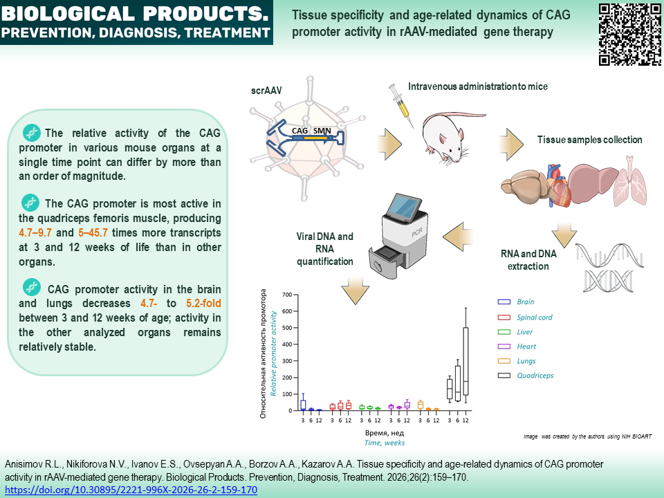

Abstract
INTRODUCTION. Artificial promoters based on the chicken beta-actin promoter (CAG, CBA, or CB) are frequently used in recombinant adeno-associated virus (rAAV)-based gene therapy products. However, data on the activity and age-related activity dynamic of these promoters remain quite limited. In this study, we quantitatively assessed CAG promoter activity in various organs of postnatal mice to determine its suitability for the development of rAAV-mediated gene therapy.
AIM. Evaluation of the relative CAG promoter activity and its dynamics in mouse organs from 3 to 12 weeks of life.
MATERIALS AND METHODS. Two-day-old ICR (CD-1) mice were injected once with rAAV9 carrying a cassette for SMN expression under the control of the CAG promoter. At 3, 6, and 12 weeks after administration, total DNA and RNA were isolated from organ samples (brain, spinal cord, liver, lungs, heart, quadriceps femoris muscle). The content of rAAV genomes and SMN mRNA in nucleic acid preparations were determined by quantitative PCR. Relative promoter activity was calculated as the ratio of the SMN mRNA concentration to the concentration of rAAV genomes, normalized on the RNA:DNA ratio in the organ. Data were analyzed using the geometric mean and regression analysis.
RESULTS. CAG promoter activity varies significantly in different organs. At week 3 after injection, the highest values were observed for the quadriceps femoris muscle, 4.7–9.7 times higher than those in other organs. It was also found that in the brain and lungs, promoter activity decreased 4.7-fold (p=0.0094) and 5.2-fold (p=0.0039), respectively, from 3 to 12 weeks after injection, while in other tissues no significant changes in activity were observed.
CONCLUSIONS. The CAG promoter is poorly suited for transgene expression in the lung and brain cells due to promoter silencing and is suboptimal for expression in the liver cells because of relatively low activity; these findings should be taken into account in the development of gene therapy products.
Keywords
Funding: The work was funded by GENERIUM JSC
About the Authors
R. L. Anisimov
GENERIUM JSC
Russian Federation
Russian Federation
Roman L. Anisimov, Cand. Sci. (Biol.)
14 Vladimirskaya St., Volginsky, Pokrov, Vladimir Region 601125
N. V. Nikiforova
GENERIUM JSC
Russian Federation
Russian Federation
Natalya V. Nikiforova
14 Vladimirskaya St., Volginsky, Pokrov, Vladimir Region 601125
E. S. Ivanov
GENERIUM JSC
Russian Federation
Russian Federation
Evgeny S. Ivanov
14 Vladimirskaya St., Volginsky, Pokrov, Vladimir Region 601125
A. A. Ovsepyan
GENERIUM JSC
Russian Federation
Russian Federation
Armen A. Ovsepyan
14 Vladimirskaya St., Volginsky, Pokrov, Vladimir Region 601125
A. A. Borzov
GENERIUM JSC
Russian Federation
Russian Federation
Anton A. Borzov
14 Vladimirskaya St., Volginsky, Pokrov, Vladimir Region 601125
A. A. Kazarov
GENERIUM JSC
Russian Federation
Russian Federation
Alexander A. Kazarov
14 Vladimirskaya St., Volginsky, Pokrov, Vladimir Region 601125
References
1. Wang J-H, Gessler DJ, Zhan W, et al. Adeno-associated virus as a delivery vector for gene therapy of human diseases. Signal Transduct Target Ther. 2024;9(1):78. https://doi.org/10.1038/s41392-024-01780-w
2. Arjomandnejad M, Dasgupta I, Flotte TR, Keeler AM. Immunogenicity of recombinant adeno-associated virus (AAV) vectors for gene transfer. BioDrugs. 2023;37(3):311–29. https://doi.org/10.1007/s40259-023-00585-7
3. Dhungel BP, Winburn I, Pereira CDF, et al. Understanding AAV vector immunogenicity: From particle to patient. Theranostics. 2024;14(3):1260–88. https://doi.org/10.7150/thno.89380
4. Choi V, McCarty D, Samulski R. AAV hybrid serotypes: Improved vectors for gene delivery. Curr Gene Ther. 2005;5(3):299–310. https://doi.org/10.2174/1566523054064968
5. Chen H. Adeno-associated virus vectors for human gene therapy. World J Med Genet. 2015;5(3):28–45. https://doi.org/10.5496/wjmg.v5.i3.28
6. Penaud-Budloo M, Le Guiner C, Nowrouzi A, et al. Adeno--associated virus vector genomes persist as episomal chromatin in primate muscle. J Virol. 2008;82(16):7875–85. https://doi.org/10.1128/JVI.00649-08
7. Muhuri M, Levy DI, Schulz M, et al. Durability of transgene expression after rAAV gene therapy. Mol Ther. 2022;30(4):1364–80. https://doi.org/10.1016/j.ymthe.2022.03.004
8. Vandamme C, Adjali O, Mingozzi F. Unraveling the complex story of immune responses to aav vectors trial after trial. Hum Gene Ther. 2017;28(11):1061–74. https://doi.org/10.1089/hum.2017.150
9. Skopenkova VV, Egorova TV, Bardina MV. Muscle-specific promoters for gene therapy. Acta Naturae. 2021;13(1):47–58. https://doi.org/10.32607/actanaturae.11063
10. Artemyev V, Gubaeva A, Paremskaia AI, et al. Synthetic promoters in gene therapy: Design approaches, features and applications. Cells. 2024;13(23):1963. https://doi.org/10.3390/cells13231963
11. Niwa H, Yamamura K, Miyazaki J. Efficient selection for high-expression transfectants with a novel eukaryotic vector. Gene. 1991;108(2):193–9. https://doi.org/10.1016/0378-1119(91)90434-d
12. O’Connor DM, Lutomski C, Jarrold MF, et al. Lot-to-lot variation in adeno-associated virus serotype 9 (AAV9) preparations. Hum Gene Ther Methods. 2019;30(6):214–25. https://doi.org/10.1089/hgtb.2019.105
13. Orbán TI, Apáti Á, Németh A, et al. Applying a “double-feature” promoter to identify cardiomyocytes differentiated from human embryonic stem cells following transposon-based gene delivery. Stem Cells. 2009;27(5):1077–87. https://doi.org/10.1002/stem.45
14. Chai S, Wakefield L, Norgard M, et al. Strong ubiquitous micro-promoters for recombinant adeno-associated viral vectors. Mol Ther Methods Clin Dev. 2023;29:504–12. https://doi.org/10.1016/j.omtm.2023.05.013
15. Fagoe ND, Eggers R, Verhaagen J, Mason MRJ. A compact dual promoter adeno-associated viral vector for efficient deli-very of two genes to dorsal root ganglion neurons. Gene Ther. 2014;21(3):242–52. https://doi.org/10.1038/gt.2013.71
16. Gray SJ, Foti SB, Schwartz JW, et al. Optimizing promoters for recombinant adeno-associated virus-mediated gene expression in the peripheral and central nervous system using self-complementary vectors. Hum Gene Ther. 2011;22(9):1143–53. https://doi.org/10.1089/hum.2010.245
17. Maguire CA, Ramirez SH, Merkel SF, et al. Gene therapy for the nervous system: Challenges and new strategies. Neurotherapeutics. 2014;11(4):817–39. https://doi.org/10.1007/s13311-014-0299-5
18. Kaspar BK, Hatfield JM, Balleydier J, et al. Means and method for producing and purifying viral vectors. US Patent No. 12168777B2; 2024.
19. Qin JY, Zhang L, Clift KL, et al. Systematic comparison of constitutive promoters and the doxycycline-inducible promoter. PLoS One. 2010;5(5):e10611. https://doi.org/10.1371/journal.pone.0010611
20. Cayer M-P, Drouin M, Sea S-P, et al. Comparison of promoter activities for efficient expression into human B cells and haematopoietic progenitors with adenovirus Ad5/F35. J Immunol Methods. 2007;322(1–2):118–27. https://doi.org/10.1016/j.jim.2007.02.008
21. Chung S, Andersson T, Sonntag K, et al. Analysis of different promoter systems for efficient transgene expression in mouse embryonic stem cell lines. Stem Cells. 2002;20(2):139–45. https://doi.org/10.1634/stemcells.20-2-139
22. Levine A, Cantoni GL, Razin A. Inhibition of promoter activi-ty by methylation: Possible involvement of protein mediators. Proc Natl Acad Sci USA. 1991;88(15):6515–8. https://doi.org/10.1073/pnas.88.15.6515
23. Rosenqvist N, Hård Af Segerstad C, Samuelsson C, et al. Activation of silenced transgene expression in neural precursor cell lines by inhibitors of histone deacetylation. J Gene Med. 2002;4(3):248–57. https://doi.org/10.1002/jgm.268
24. Zhang F, Frost AR, Blundell MP, et al. A ubiquitous chromatin opening element (UCOE) confers resistance to DNA methylation-mediated silencing of lentiviral vectors. Mol Ther. 2010;18(9):1640–9. https://doi.org/10.1038/mt.2010.132
25. Schuster DJ, Dykstra JA, Riedl MS, et al. Biodistribution of adeno-associated virus serotype 9 (AAV9) vector after intrathecal and intravenous delivery in mouse. Front Neuroanat. 2014;8:42. https://doi.org/10.3389/fnana.2014.00042
26. Dayton RD, Wang DB, Klein RL. The advent of AAV9 expands applications for brain and spinal cord gene delivery. Expert Opin Biol Ther. 2012;12(6):757–66. https://doi.org/10.1517/14712598.2012.681463
27. Mohan RR, Schultz GS, Hong J-W, et al. Gene transfer into rabbit keratocytes using AAV and lipid-mediated plasmid DNA vectors with a lamellar flap for stromal access. Exp Eye Res. 2003;76(3):373–83. https://doi.org/10.1016/S0014-4835(02)00275-0
28. Ma H, Bell KN, Loker RN. qPCR and qRT-PCR analysis: Regulatory points to consider when conducting biodistribution and vector shedding studies. Mol Ther Methods Clin Dev. 2020;20:152–68. https://doi.org/10.1016/j.omtm.2020.11.007
29. Bustin S. Absolute quantification of mRNA using real-time reverse transcription polymerase chain reaction assays. J Mol Endocrinol. 2000;25(2):169–93. https://doi.org/10.1677/jme.0.0250169
30. Schmidt EE, Schibler U. Cell size regulation, a mechanism that controls cellular RNA accumulation: Consequences on regulation of the ubiquitous transcription factors Oct1 and NF-Y and the liver-enriched transcription factor DBP. J Cell Biol. 1995;128(4):467–83. https://doi.org/10.1083/jcb.128.4.467
31. McCarty DM, Fu H, Monahan PE, et al. Adeno-associated virus terminal repeat (TR) mutant generates self-complementary vectors to overcome the rate-limiting step to transduction in vivo. Gene Ther. 2003;10(26):2112–8. https://doi.org/10.1038/sj.gt.3302134
32. Xiao X, Li J, Samulski RJ. Production of high-titer recombinant adeno-associated virus vectors in the absence of helper adenovirus. J Virol. 1998;72(3):2224–32. https://doi.org/10.1128/JVI.72.3.2224-2232.1998
33. Xu J, DeVries SH, Zhu Y. Quantification of adeno-associated virus with safe nucleic acid dyes. Hum Gene Ther. 2020; 31(19–20):1086–99. https://doi.org/10.1089/hum.2020.063
34. Fowler WM, Taylor RG, Franti CE, Hagler AN. The effect of age on the nucleic acid content of slow- and fast-twitch muscle in normal and dystrophic mice and their litter mates. J Neurol Sci. 1977;32(2):227–41. https://doi.org/10.1016/0022-510x(77)90238-6
35. Keinath MC, Prior DE, Prior TW. Spinal muscular atrophy: Mutations, testing, and clinical relevance. Appl Clin Genet. 2021;14:11–25. https://doi.org/10.2147/TACG.S239603
36. Mendell JR, Al-Zaidy S, Shell R, et al. Single-dose gene-replacement therapy for spinal muscular atrophy. N Engl J Med. 2017;377(18):1713–22. https://doi.org/10.1056/NEJMoa1706198
37. Rhee J, Kang J, Jo Y, et al. Improved therapeutic approach for spinal muscular atrophy via ubiquitination-resistant survival motor neuron variant. J Cachexia Sarcopenia Muscle. 2024;15(4):1404–17. https://doi.org/10.1002/jcsm.13486
38. Cearley CN, Wolfe JH. Transduction characteristics of adeno-associated virus vectors expressing cap serotypes 7, 8, 9, and Rh10 in the mouse brain. Mol Ther. 2006;13(3):528–37. https://doi.org/10.1016/j.ymthe.2005.11.015
39. Gray SJ, Matagne V, Bachaboina L, et al. Preclinical differences of intravascular AAV9 delivery to neurons and glia: A comparative study of adult mice and nonhuman primates. Mol Ther. 2011;19(6):1058–69. https://doi.org/10.1038/mt.2011.72
40. Miyake N, Miyake K, Yamamoto M, et al. Global gene transfer into the CNS across the BBB after neonatal systemic delivery of single-stranded AAV vectors. Brain Res. 2011;1389:19–26. https://doi.org/10.1016/j.brainres.2011.03.014
41. Bevan AK, Duque S, Foust KD, et al. Systemic gene delivery in large species for targeting spinal cord, brain, and peripheral tissues for pediatric disorders. Mol Ther. 2011;19(11):1971–80. https://doi.org/10.1038/mt.2011.157
42. Hammelrath L, Škokić S, Khmelinskii A, et al. Morphological maturation of the mouse brain: An in vivo MRI and histology investigation. Neuroimage. 2016;125:144–52. https://doi.org/10.1016/j.neuroimage.2015.10.009
43. Cisternas CD, Cortes LR, Bruggeman EC, et al. Developmental changes and sex differences in DNA methylation and demethylation in hypothalamic regions of the mouse brain. Epigenetics. 2020;15(1–2):72–84. https://doi.org/10.1080/15592294.2019.1649528
44. Lister R, Mukamel EA, Nery JR, et al. Global epigenomic reconfiguration during mammalian brain development. Science. 2013;341(6146):1237905. https://doi.org/10.1126/science.1237905
45. Ellis H. Anatomy of head injury. Surg Oxf. 2007;25:505–7. https://doi.org/10.1016/j.mpsur.2007.10.008
46. Miyanohara A, Kamizato K, Juhas S, et al. Potent spinal parenchymal AAV9-mediated gene delivery by subpial injection in adult rats and pigs. Mol Ther Methods Clin Dev. 2016;3:16046. https://doi.org/10.1038/mtm.2016.46
47. Duque S, Joussemet B, Riviere C, et al. Intravenous administration of self-complementary AAV9 enables transgene delivery to adult motor neurons. Mol Ther. 2009;17(7):1187–96. https://doi.org/10.1038/mt.2009.71
48. Chand D, Mohr F, McMillan H, et al. Hepatotoxicity following administration of onasemnogene abeparvovec (AVXS-101) for the treatment of spinal muscular atrophy. J Hepatol. 2021;74(3):560–6. https://doi.org/10.1016/j.jhep.2020.11.001
49. Khan SA, Álvarez JV, Nidhi FNU, et al. Evaluation of AAV vectors with tissue-specific or ubiquitous promoters in a mouse model of mucopolysaccharidosis type IVA. Mol Ther Methods Clin Dev. 2025;33(2):101447. https://doi.org/10.1016/j.omtm.2025.101447
50. Schittny JC, Mund SI, Stampanoni M. Evidence and structural mechanism for late lung alveolarization. Am J Physiol Lung Cell Mol Physiol. 2008;294(2):L246–54. https://doi.org/10.1152/ajplung.00296.2007
51. Mund SI, Stampanoni M, Schittny JC. Developmental alveolarization of the mouse lung. Dev Dyn. 2008;237(8):2108–16. https://doi.org/10.1002/dvdy.21633
52. Tawa R, Ono T, Kurishita A, et al. Changes of DNA methylation level during pre- and postnatal periods in mice. Differentiation. 1990;45(1):44–8. https://doi.org/10.1111/j.1432-0436.1990.tb00455.x
53. Cuna A, Halloran B, Faye-Petersen O, et al. Alterations in gene expression and DNA methylation during murine and human lung alveolar septation. Am J Respir Cell Mol Biol. 2015;53(1):60–73. https://doi.org/10.1165/rcmb.2014-0160OC
54. Spalding KL, Bhardwaj RD, Buchholz BA, et al. Retrospective birth dating of cells in humans. Cell. 2005;122(1):133–43. https://doi.org/10.1016/j.cell.2005.04.028
55. Wang Z, Zhu T, Qiao C, et al. Adeno-associated virus serotype 8 efficiently delivers genes to muscle and heart. Nat Biotechnol. 2005;23(3):321–8. https://doi.org/10.1038/nbt1073
56. White RB, Biérinx A-S, Gnocchi VF, Zammit PS. Dynamics of muscle fibre growth during postnatal mouse development. BMC Dev Biol. 2010;10:21. https://doi.org/10.1186/1471-213X-10-21
57. Leu M, Ehler E, Perriard J-C. Characterisation of postnatal growth of the murine heart. Anat Embryol (Berl). 2001;204(3):217–24. https://doi.org/10.1007/s004290100206
58. Swift SK, Purdy AL, Kolell ME, et al. Cardiomyocyte ploidy is dynamic during postnatal development and varies across genetic backgrounds. Development. 2023;150(7):dev201318. https://doi.org/10.1242/dev.201318
59. Borowik AK, Davidyan A, Peelor FF, et al. Skeletal muscle nuclei in mice are not post-mitotic. Function (Oxf). 2022;4(1):zqac059. https://doi.org/10.1093/function/zqac059
60. Simon A, Djeddi S, Bournon P, et al. Transcriptomic characterization of postnatal muscle maturation. Dis Model Mech. 2025;18(2):DMM052098. https://doi.org/10.1242/dmm.052098
61. Stieger K, Schroeder J, Provost N, et al. Detection of intact rAAV particles up to 6 years after successful gene transfer in the retina of dogs and primates. Mol Ther. 2009;17(3):516–23. https://doi.org/10.1038/mt.2008.283
62. Stone D, Liu Y, Li Z-Y, et al. Biodistribution and safety profile of recombinant adeno-associated virus serotype 6 vectors following intravenous delivery. J Virol. 2008;82(15):7711–5. https://doi.org/10.1128/JVI.00542-08
63. Munguía-Fuentes R, Yam-Puc JC, Silva-Sánchez A, et al. Immunization of newborn mice accelerates the architectural maturation of lymph nodes, but AID-dependent IgG responses are still delayed compared to the adult. Front Immunol. 2017;8:13. https://doi.org/10.3389/fimmu.2017.00013
64. Massaro G, Geard AF, Nelvagal HR, et al. Comparison of different promoters to improve AAV vector-mediated gene therapy for neuronopathic Gaucher disease. Hum Mol Genet. 2024;33(17):1467–80. https://doi.org/10.1093/hmg/ddae081
65. King AD, Huang K, Rubbi L, et al. Reversible regulation of promoter and enhancer histone landscape by DNA methylation in mouse embryonic stem cells. Cell Rep. 2016;17(1):289–302. https://doi.org/10.1016/j.celrep.2016.08.083
66. Ryczek N, Łyś A, Makałowska I. The functional meaning of 5’UTR in protein-coding genes. Int J Mol Sci. 2023;24(3):2976. https://doi.org/10.3390/ijms24032976
67. Garza IT, Eller MM, Holmes SK, et al. Expression and distribution of rAAV9 intrathecally administered in juvenile to adolescent mice. Gene Ther. 2025;32(3):189–96. https://doi.org/10.1038/s41434-024-00498-2
68. Sinturel F, Gerber A, Mauvoisin D, et al. Diurnal oscillations in liver mass and cell size accompany ribosome assembly cycles. Cell. 2017;169(4):651–63.e14. https://doi.org/10.1016/j.cell.2017.04.015
Supplementary files

|
1. Supplementary material 1 | |
| Subject | ||
| Type | Исследовательские инструменты | |
Download
(844KB)
|
Indexing metadata ▾ | |
|
|
2. Supplementary material 2 | |
| Subject | ||
| Type | Исследовательские инструменты | |
Download
(747KB)
|
Indexing metadata ▾ | |
Review
For citations:
Anisimov R.L., Nikiforova N.V., Ivanov E.S., Ovsepyan A.A., Borzov A.A., Kazarov A.A. Tissue specificity and age-related dynamics of CAG promoter activity in rAAV-mediated gene therapy. Biological Products. Prevention, Diagnosis, Treatment. 2026;26(2):159-170. (In Russ.) https://doi.org/10.30895/2221-996X-2026-26-2-159-170
Views:
836
JATS XML





 Email this article