



Содержание
Перейти к:
https://doi.org/10.30895/2221-996X-2026-26-2-159-170
Перейти к:
ВВЕДЕНИЕ. Несмотря на широкое использование в генотерапевтических препаратах на основе рекомбинантных аденоассоциированных вирусов (rAAV) искусственных промоторов, основанных на промоторе β-актина курицы (обозначаемых как CAG, CBA или CB), данные об активности таких промоторов в разных тканях и возрастной динамике остаются ограниченными. В настоящей работе проведена количественная оценка активности CAG-промотора в различных органах мышей в постнатальном периоде для определения его применимости при разработке rAAV-опосредованной терапии.
ЦЕЛЬ. Оценка относительной активности CAG-промотора и ее динамики в органах мышей в возрасте 3–12 недель.
МАТЕРИАЛЫ И МЕТОДЫ. 2-дневным мышам линии ICR (CD-1) однократно вводили rAAV9 с кассетой для экспрессии SMN под контролем CAG-промотора. На 3, 6 и 12-й нед. после инъекции из образцов органов (головной мозг, спинной мозг, печень, легкие, сердце, четырехглавая мышца бедра) выделяли тотальную ДНК и РНК. Содержание вирусных геномов rAAV и мРНК SMN в препаратах нуклеиновых кислот определяли методом количественной ПЦР. Относительную активность промотора рассчитывали как отношение концентрации мРНК SMN к концентрации вирусных геномов (rAAV), нормализованное на соотношение РНК:ДНК в органе. Для анализа данных вычисляли среднее геометрическое и использовали регрессионный анализ.
РЕЗУЛЬТАТЫ. Активность CAG-промотора значительно варьировала в разных органах. На 3 нед. после инъекции наибольшие значения активности отмечены в четырехглавой мышце бедра, в 4,7–9,7 раза превышающие таковые в других органах. Также обнаружено, что в головном мозге и легких с 3 по 12 нед. после инъекции активность промотора снижалась в 4,7 раза (p=0,0094) и в 5,2 раза (p=0,0039) соответственно, в то время как в других тканях существенных изменений активности не наблюдалось.
ВЫВОДЫ. CAG-промотор малопригоден для экспрессии трансгена в клетках легких и головного мозга вследствие транскрипционного сайленсинга, а также не является оптимальным для экспрессии в клетках печени из-за относительно низкой активности. Эти данные следует учитывать при разработке генотерапевтических препаратов.
Анисимов Р.Л., Никифорова Н.В., Иванов Е.С., Овсепян А.А., Борзов А.А., Казаров А.А. Активность CAG-промотора при rAAV-опосредованной генной терапии: тканеспецифичность и возрастная динамика. БИОпрепараты. Профилактика, диагностика, лечение. 2026;26(2):159-170. https://doi.org/10.30895/2221-996X-2026-26-2-159-170
Anisimov R.L., Nikiforova N.V., Ivanov E.S., Ovsepyan A.A., Borzov A.A., Kazarov A.A. Tissue specificity and age-related dynamics of CAG promoter activity in rAAV-mediated gene therapy. Biological Products. Prevention, Diagnosis, Treatment. 2026;26(2):159-170. (In Russ.) https://doi.org/10.30895/2221-996X-2026-26-2-159-170
Рекомбинантные аденоассоциированные вирусы (recombinant adeno-associated viruses, rAAV) в настоящее время из высокотехнологичного инструмента превращаются в один из базовых методов генной терапии. В настоящее время зарегистрировано несколько препаратов на основе rAAV, применяемых для терапии орфанных заболеваний (семейная недостаточность липопротеинлипазы, наследственная дистрофия сетчатки, гемофилия B) [1]. rAAV считаются относительно малоиммуногенными [2][3], инфицируют широкий спектр клеток [4] и относительно просты в производстве [5]. Геном rAAV существует в клетке хозяина в виде нереплицирующихся эписом [6], поэтому применение rAAV для устойчивого терапевтического эффекта ограничено долгоживущими и медленно делящимися клетками [7], а возможность системного введения лимитирована возникновением иммунного ответа [8].
Геном rAAV представляет собой экспрессионную кассету, включающую промотор, кодирующую последовательность и сайт полиаденилирования, фланкированную инвертированными повторами (inverted terminal repeats, ITR). Промоторы условно разделяют на тканеспецифичные (например, производные промотора мышечной креатинкиназы, широко используемые в конструкциях для генной терапии наследственных заболеваний мышц [9]) и промоторы широкого спектра, позволяющие экспрессировать трансген в клетках практически любых тканей (например, промоторы PGK, CMV, EF1a) [10]. Одними из самых популярных промоторов в клинических исследованиях являются укороченные производные искусственного CAG‑промотора, состоящего из раннего энхансера цитомегаловируса, промотора, первого интрона и экзона β-актина курицы, а также акцептора сплайсинга β-глобина кролика [11]. Поскольку исходный промотор имеет очень большие размеры (около 1,7 тыс. п.н.), обычно используются его укороченные варианты, в которых неизменной остается только коровая часть промотора β-актина. Эти производные обозначаются в литературе как «CAG» [12–15], «CBA» (chicken beta actin) [16] и «CB» [17]. Такой промотор, состоящий из промотора β-актина курицы, энхансера цитомегаловируса и модифицированного позднего 16S интрона вируса SV40, используется, например, в экспрессионной конструкции препарата Золгенсма (онасемноген абепарвовек, Novartis), применяемого для терапии спинальной мышечной атрофии [18].
Однако в действительности широкая специфичность промотеров не абсолютна, их активность сильно зависит от видовой принадлежности и типа клеток [19]. Например, промотор CMV, часто используемый для экспрессии трансгена в клетках HEK293 и CHO, имеет низкую активность в B-лимфоцитах [20] и практически полностью инактивируется в эмбриональных стволовых клетках мыши [21]. Кроме того, активность промоторов в разных тканях может по-разному изменяться со временем из-за влияния эпигенетических факторов (CpG-метилирование, модификация гистонов) [22–24].
Эти особенности промоторов необходимо учитывать при разработке генетической конструкции генотерапевтических препаратов, так как обеспечение достаточного уровня экспрессии трансгена в целевой ткани в течение долгого времени определяет эффективность, безопасность и стоимость препарата. Однако, несмотря на объективную необходимость оценки активности промоторов и ее динамики в различных органах и тканях, имеющиеся данные весьма ограничены.
Исследователи, тестирующие новые варианты rAAV или способы их введения, часто ограничиваются только определением уровня экспрессии репортерного гена (зеленый флуоресцентный белок (GFP), β-галактозидаза или хлорамфениколацетилтрансфераза) в целевых органах и клетках [16][25–27]. Такой подход достаточен для целей исследований, но не дает информации об активности промотора, поскольку уровень экспрессии зависит также от эффективности вирусной трансдукции клеток органа.
Более информативен подход, рекомендуемый ведущими регуляторными органами (Европейское агентство по лекарственным средствам и Управление по контролю за качеством продуктов питания и лекарственных средств США), в котором для оценки биораспределения вектора и экспрессии трансгена используется количественная полимеразная цепная реакция в реальном времени (ПЦР-РВ) и количественная полимеразная цепная реакция с обратной транскрипцией (ОТ-ПЦР) [28]. В таких исследованиях применяют нормализацию на единицу массы тотальной ДНК и РНК [28][29]. Располагая данными о количестве вирусной ДНК и РНК трансгена в органах, можно оценить относительную активность промоторов, что позволяет прогнозировать результаты исследований с использованием тех же промоторов в экспрессионных кассетах rAAV. Однако при этом не учитывается, что количество РНК в клетках организма может варьировать более чем в 16 раз в зависимости от органа [30]. Соответственно, при теоретически одинаковой активности промотора для печени, где соотношение РНК:ДНК максимально среди органов, измеряемые значения активности будут занижены в 3 раза по сравнению с таковыми в сердце и в 8–9 раз — в легких. Таким образом, для корректного расчета относительной активности промотора в разных органах следует вводить поправочный коэффициент, отражающий относительное содержание РНК, то есть соотношение РНК:ДНК для каждого органа.
Цель работы — оценка относительной активности CAG‑промотора и ее динамики в органах мышей в возрасте 3–12 недель.
Фланкированная ITR экспрессионная кассета (Последовательность 1, опубликована в Приложении 1 на сайте журнала1), содержащая последовательность гибридного CAG‑промотора, оптимизированную для экспрессии в клетках млекопитающих кодирующую последовательность белка SMN (survival motor neuron) и сайт полиаденилирования SV40, была синтезирована в TOP Gene Technologies (Канада). В последовательности одного из ITR была делетирована область trs (терминальный сайт расщепления, terminal resolution site) для получения scrAAV (самокомплементарный рекомбинантный AAV, self-complementary recombinant AAV) [31]. Последовательность CAG‑промотора была аналогична использовавшейся в оригинальном препарате (Золгенсма, Novartis) [18], с измененными сайтами клонирования между элементами промотора и добавленной канонической последовательностью Козак ACCGCCACCATG. Полученная последовательность была клонирована в вектор, производный от pUС, с геном устойчивости к канамицину для получения rAAV-плазмиды pAAV-CAG‑SMN.
Плазмида pT7-SMN для наработки РНК SMN с помощью Т7 РНК-полимеразы была создана путем замены последовательности энхансера цитомегаловируса и промотора β-актина курицы в плазмиде pAAV-CAG‑SMN1 на последовательность Т7 промотора TAATACGACTCACTATAGGG.
Наработку вирусов проводили по традиционной методике [32] c использованием трех плазмид: pHelper (GenBank AF369965.1), pRep2Cap9 (кат. № 112865, Addgene) и pAAV-CAG‑SMN, которыми в эквимолярных соотношениях одновременно трансфицировали клетки HEK293. Вирусы выделяли из клеточного лизата с помощью последовательных хроматографических очисток на сорбентах POROS Capture Select AAV9 и POROS 50 HQ (Thermo Fisher Scientific, США) согласно инструкции производителя. Полученный препарат анализировали на содержание остаточных белков хозяина (менее 10 нг/мл) и концентрировали до 2,0×10¹³ вирусных геномов/мл в буфере готовой лекарственной формы (ГЛФ) (200 мМ NaCl, 2 мМ MgCl2, 20 мМ трис(гидроксиметил)аминометана, 0,005% Poloxamer 188 (Pluronic F68), pH 8,0). Заполненность капсидов оценивали методом аналитического ультрацентрифугирования (≈97,2%). Целостность и самокомплементарность ДНК рекомбинантного вируса подтверждены методом щелочного электрофореза [33].
Мыши линии ICR CD-1 обоего пола были получены из НПП «Питомник лабораторных животных» (Филиал ФГБУН «Государственный научный центр Институт биоорганической химии им. академиков М.М. Шемякина и Ю.А. Овчинникова» РАН, Пущино, Московская обл.). Перед введением в исследование животные, из которых впоследствии формировались родительские пары, проходили адаптацию в течение 8 сут. Содержание и манипуляции с животными проводили в соответствии с требованиями Международной конвенции по защите позвоночных животных, используемых для экспериментальных и научных целей2, а также Директивы 2010/63/EU Европейского парламента и Совета Европейского союза по охране животных, используемых в научных целях3. Протокол исследования рассмотрен и утвержден на заседании Комиссии по биоэтике АО «ГЕНЕРИУМ» (ветеринарный протокол № 64, версия 1 от 05.08.2022).
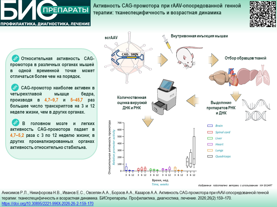
Для изучения активности CAG‑промотора в органах использовались новорожденные 2-дневные мыши линии CD-1 (n=20) в соответствии с утвержденной схемой исследования (рис. 1; раздел «Схема исследований», Приложение 1) и с учетом данных доклинических исследований оригинального препарата4. Работа с экспериментальными животными описана в разделе «Манипуляции с экспериментальными животными» Приложения 1.

Рисунок подготовлен авторами с использованием ресурса NIH BIOART5 / The figure was prepared by the authors using the NIH BIOART Source5
Рис. 1. Схема исследования. scrAAV — самокомплементарный рекомбинантный AAV.
Fig. 1. Experimental design. scrAAV, self-complementary recombinant AAV.
Для выделения нуклеиновых кислот забирали по три образца тканей сердца, легких, печени, мозга и четырехглавой мышцы бедра (квадрицепса) массой 10±2 мг, а также спинной мозг (целиком). Процедура детально описана в разделе «Забор образцов тканей и выделение нуклеиновых кислот» Приложения 1.
Оценка количества геномов rAAV в тканях была проведена методом количественной ПЦР в реальном времени (ПЦР-РВ) с использованием системы CFX96 Touch real-time PCR detection system (Bio-Rad, США). Условия проведения ПЦР-РВ приведены в разделе «Количественная оценка вирусной ДНК и мРНК в образцах тканей» Приложения 1 и таблицах S1–S4 Приложения 1.
Относительную активность промотора рассчитывали как отношение числа транскриптов SMN в 25 нг тотальной РНК к числу копий вирусных геномов в 50 нг тотальной ДНК для каждого образца, умноженное на коэффициент РНК:ДНК (табл. S5, Приложение 1). Данные о соотношении РНК:ДНК для головного мозга, печени, легких и сердца взяты из работы E.E. Schmidt и U. Schibler [30]; для спинного мозга использовались те же значения, что и для головного мозга. Для четырехглавой мышцы бедра использовали усредненные значения, полученные W.M. Fowler с соавт. [34] для икроножной мышцы мышей в возрасте 4–13 нед. (табл. S5, Приложение 1).
Все процедуры статистического анализа выполняли с использованием программного обеспечения GraphPad Prism 9.5 (GraphPad Software Inc., США). Распределение значений содержания векторной ДНК, мРНК-транскриптов SMN и относительной активности CAG‑промотора статистически значимо отличалось от нормального и соответствовало лог-нормальному распределению (критерии Шапиро — Уилка и д’Агостино — Пирсона). В связи с этим для оценки математического ожидания и размаха данных использовали геометрическое среднее (GM) и геометрический коэффициент вариации (GCV, %) (табл. S6–S8 опубликованы в Приложении 2 на сайте журнала6). Для оценки временной динамики содержания векторной ДНК и относительной активности промотора (после логарифмического преобразования по основанию 10 для приведения данных к нормальному распределению) применяли метод линейного регрессионного анализа. Для оценки средней скорости изменения показателей (Δ, % в нед.) использовали формулу (1):
Δ = 100% × (10k – 1), (1)
где k — угловой коэффициент линейной зависимости логарифма показателя от времени.
По количеству вирусных геномов в 50 нг тотальной ДНК, выделенной из образцов органов мышей в возрасте 3 нед., органы можно разделить на две категории: с условно низким содержанием (до 6000 копий) — головной мозг, спинной мозг и печень; с высоким содержанием (более 20000 копий) — легкие, сердце, четырехглавая мышца бедра (рис. 2; табл. S6 Приложение 2). Наименьшее содержание было отмечено в ДНК, выделенной из спинного мозга (1863 копии, GCV 102%), а наибольшее — в препарате из сердца (42365 копий, GCV 34%).
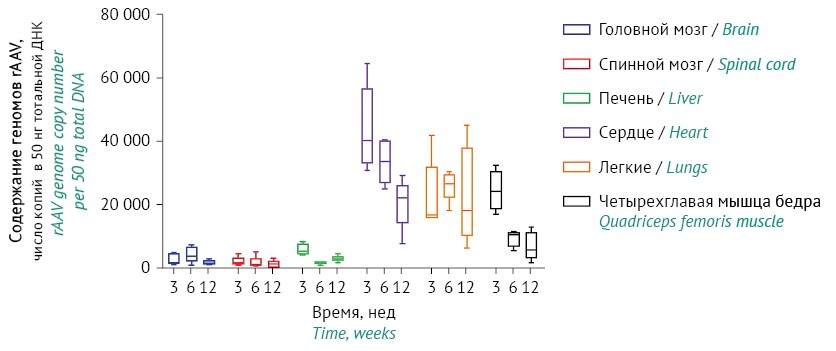
Рисунок подготовлен авторами по собственным данным / The figure was prepared by the authors using their own data
Рис. 2. Число копий геномов rAAV в 50 нг тотальной ДНК органов мышей.
Fig. 2. rAAV genome copy number in 50 ng of total DNA from mouse organs.
С течением времени содержание вирусных геномов изменялось неравномерно (табл. S9, Приложение 2). Так, в головном мозге и легких оно находилось примерно на одном уровне; в печени наблюдалось более чем двукратное снижение с 3 по 6 нед. с дальнейшим выходом на плато; для остальных органов происходило снижение в 2–3 раза с 3 по 12 нед., наиболее выраженное для препаратов из четырехглавой мышцы (с GM 23800 до 5609 копий, p=0,0001).
Для головного и спинного мозга отмечена наибольшая вариативность: содержание геномов rAAV для разных животных в одной временной точке могло различаться более чем на порядок.
Активность промотора сильно различалась в органах. Наименьшая активность промотора у мышей в возрасте 3 нед. наблюдалась в головном мозге (11,85), а наибольшая — в четырехглавой мышце (115,33), в 4,7–9,7 раза превышающая активность в других органах (рис. 3; табл. S8, Приложение 2). В головном мозге также отмечена наибольшая вариативность (GCV 348%). Активность промотора для части проб менялась со временем как в сторону увеличения, так и уменьшения (табл. S10, Приложение 2). По этому признаку органы можно разделить на две условные группы: в одной активность промотора с 3 по 12 нед. значимо снижалась в несколько раз (головной мозг p=0,0094; легкие p=0,0039), в другой оставалась приблизительно постоянной (печень, сердце) или умеренно повышалась (спинной мозг р>0,05; четырехглавая мышца р=0,0226). Так, активность промотора в головном мозге снизилась в 4,7 раза (с 11,85 до 2,52), в то время как в четырехглавой мышце бедра увеличилась на 68% (со 115,33 до 194,24).

Рисунок подготовлен авторами по собственным данным / The figure was prepared by the authors using their own data
Рис. 3. Относительная активность CAG‑промотора в органах мышей.
Fig. 3. Relative activity of the CAG promoter in mouse organs.
В проведенном исследовании использовался rAAV, аналогичный таковому в оригинальном препарате (Золгенсма), предназначенном для лечения спинальной мышечной атрофии. При этом заболевании из-за мутаций в гене SMN1 происходит дегенерация двигательных нейронов спинного мозга в постнатальный период [35]. Предполагается, что оригинальный препарат, представляющий собой rAAV9, содержащий кассету для экспрессии SMN, восполняет мутированный ген. Однако в клинических испытаниях вводимый внутривенно препарат показал низкую эффективность [36][37]. Предположительно, одной из причин является неудачный выбор промотора экспрессионной кассеты и способа введения, приводящие к повышенной вирусной нагрузке в нецелевых органах и низкой экспрессии в целевых. В представленном исследовании проведена оценка возможности применения CAG‑промотора для экспрессии трансгена в различных органах и тканях.
Головной и спинной мозг. Серотип AAV9 часто используется для генной терапии благодаря способности проникать через гематоэнцефалический барьер и заражать клетки центральной нервной системы [26][38][39], но он так же эффективно трансдуцирует клетки тканей других органов, в частности сердца, печени и скелетных мышц [40][41]. Из полученных нами данных следует, что головной и спинной мозг не являются предпочтительной целью для rAAV9 у новорожденных мышей.
Среднее содержание геномов rAAV (GM) в головном мозге оставалось постоянным в течение всего эксперимента и снижалось примерно вдвое в образцах спинного мозга. Это можно объяснить тем, что объем головного мозга мышей, а соответственно, и количество клеток после 3-недельного возраста увеличивается приблизительно лишь на 10% [42], объем же других органов, в том числе спинного мозга, увеличивается пропорционально массе тела мышей, которая в течение эксперимента возросла в 1,5–2 раза.
Средняя активность CAG‑промотора в головном и спинном мозге у мышей 3-недельного возраста находилась на самом низком уровне среди образцов проанализированных органов. В дальнейшем активность CAG‑промотора в головном мозге уменьшалась в 4,7 раза, что, возможно, связано с изменением профиля метилирования ДНК в первые 4 нед. жизни [43][44], которое теоретически способно вызывать наблюдаемое снижение активности промотора.
Для головного и спинного мозга, несмотря на общность происхождения и непосредственную связь, отмечена резко различающаяся динамика активности CAG‑промотора: в спинном мозге измеряемая активность не только не уменьшалась, но даже увеличилась на 67%. Предположительно, разница в динамике активности обусловлена различным клеточным составом органа. Известно, что AAV9 способен инфицировать как нейроны, так и глиальные клетки [39], общие для обоих органов. Однако отбираемые образцы спинного мозга также содержали фибробласты и эндотелиоциты мягкой оболочки спинного мозга (pia mater) [45], которые являются значительно более доступной мишенью для AAV9, поскольку именно pia mater представляет собой основной барьер для проникновения AAV в паренхиму спинного мозга [46]. Поэтому, возможно, ассоциация относительно высокого уровня трансдукции клеток спинного мозга rAAV9 и последующей экспрессии трансгена, наблюдаемой во многих исследованиях [26][47], только с нейронами несостоятельна. Преимущественная трансдукция ненейронных клеток могла бы объяснить низкую эффективность оригинального препарата.
Печень. Ранее была отмечена высокая гепатотоксичность оригинального препарата Золгенсма, предположительно из-за иммунного ответа на вирусные капсиды [48]. Однако в нашем исследовании вирусная нагрузка клеток печени оказалась значительно ниже, чем в мышцах и легких, и сравнима с таковой в клетках ЦНС. Скорее всего, наблюдаемое нами низкое содержание вирусных геномов связано с выбранным способом введения (в лицевую вену), который не предполагает первоначальной фильтрации в печени, в отличие от традиционной инъекции в периферические вены (конечностей и хвоста), при которых наибольшее содержание вирусных геномов обнаруживается именно в печени, как, например, в работе S.A. Khan с соавт. [49].
Кроме того, относительно постоянная активность CAG‑промотора в печени у мышей 3-недельного возраста лишь незначительно превышает таковую в головном мозге. Таким образом, CAG‑промотор не является оптимальным для экспрессии трансгена в клетках печени из-за относительно низкой активности. Подбор более эффективного промотора позволит существенно снизить вирусную нагрузку на орган при генной терапии, направленной на печень.
Легкие. Легкие, так же как сердце и скелетные мышцы, являются преимущественной мишенью AAV9: относительное содержание ДНК rAAV в этих органах было значительно выше, чем в других исследованных органах. Количество вирусных геномов в легочной ткани варьировало в широких пределах у экспериментальных животных, но среднее содержание с 3 по 12 нед. менялось незначительно (табл. S6, Приложение 2). Однако, как и в случае головного мозга, наблюдалось снижение активности промотора с 3 по 6 нед. почти в 5 раз, с последующей стабилизацией на приблизительно постоянном уровне. Возможно, это связано с тем, что в легких к концу 3 нед. завершается активная стадия альвеоляризации и начинается формирование взрослой структуры [50][51]. Этот процесс сопровождается увеличением метилирования ДНК, приводящим к изменению экспрессии ряда генов, связанных с развитием легких [52][53]. Возможно, при этом также метилируется CAG‑промотор, что и вызывает резкое снижение его активности с 3 по 6 нед.
Сердце и четырехглавая мышца бедра. Среди проанализированных тканей именно в квадрицепсе CAG‑промотор наиболее активен, продуцируя к 12 нед. в 16 и 77 раз больше транскриптов, чем в печени или головном мозге соответственно. При отсутствии мышечно-специфических промоторов, основанных на промоторах генов креатинкиназы или десмина [9], или невозможности их использования CAG‑промотор может обеспечить высокий уровень экспрессии трансгена в мышечной ткани.
Считается, что мышечная ткань является подходящей мишенью для rAAV благодаря стабильности: средний возраст миоцитов человека составляет около 15 лет [54]. Соответственно, снижение содержания существующих в виде эписом вирусных геномов из-за деления клеток происходит очень медленно, что обеспечивает большую устойчивость (durability) экспрессии трансгена [7]. Однако нами было обнаружено значительное снижение содержания геномов rAAV в образцах квадрицепса (в 4 раза) и сердца (в 2 раза) с 3 по 12 нед. после инъекции при увеличивающемся уровне экспрессии на 68 и 29% соответственно. Полученные результаты противоречат ранее опубликованным заключениям Z. Wang с соавт. [55] о стабильном числе копий вирусного генома в мышцах. Однако следует отметить, что в упомянутой работе не приведено сведений о динамике и представлены данные по биораспределению только для одной временной точки (2 месяца после введения вируса). Возможно, наблюдаемая нами потеря геномов rAAV имеет место только в мышечных волокнах и кардиомиоцитах в стадии гипертрофии; с возрастом число вирусных геномов в этих тканях стабилизируется. В пользу этого говорит замедление снижения среднего содержания геномов rAAV с возрастом животного: изменение, которое происходит с 6 по 12 нед., сопоставимо с периодом с 3 по 6 нед.
Наблюдаемое снижение содержания геномов rAAV не может быть объяснено процессами деления мышечной ткани. Известно, что период созревания мышечных волокон, сопровождающийся увеличением числа ядер в миофибрилле, завершается к 3 нед., а дальнейший рост мышечного волокна происходит только за счет гипертрофии [56]. В сердечной мышце в экспериментальный период стадия активного деления кардиомиоцитов (1–15 сут жизни) уже завершилась, и происходит плавное увеличение размеров сердечной мышцы за счет гипертрофии кардиомиоцитов, коррелирующее с увеличением массы тела [57]. Возрастное увеличение плоидности в 3-недельном возрасте [58][59] также слишком незначительно, чтобы обеспечить наблюдаемое снижение.
Увеличение активности CAG‑промотора в мышечной ткани также труднообъяснимо. Как указано выше, стадии активных изменений в мышцах и сердце в экспериментальный период завершились, и логично было бы ожидать стабильного уровня экспрессии. Единственное значительное изменение, происходящее в скелетных мышцах, — появление специализации мышечных волокон, выражающееся в разделении их на окислительные («медленные») и гликолитические («быстрые»), начинающееся как минимум с 2-недельного возраста [60]. Возможно, увеличение активности CAG‑промотора может быть связано с этим процессом.
Еще одним объяснением как снижения измеряемого числа вирусных геномов, так и роста измеряемой активности CAG‑промотора в мышцах может быть присутствие в мышечной ткани транскрипционно неактивных вирусных частиц после инъекции. Известно, что в иммунопривилегированных тканях интактные вирусные частицы могут обнаруживаться в течение как минимум 6 лет [61]. В прочих тканях срок жизни вирусных частиц намного меньше, так как воспалительная реакция в ответ на присутствие вирусных капсидов разных серотипов полностью завершается у взрослых мышей уже в течение 72 ч [62]. Иммунная система новорожденных мышей отличается от таковой у взрослых [63], и, возможно, элиминация вирусных частиц в мышечной ткани происходит медленнее. Если вирусные частицы присутствуют в мышечной ткани в количествах, сравнимых с количеством трансдуцированных вирусных геномов или превышающих его, их элиминация ведет к увеличению измеряемой активности промотора даже при снижении относительного содержания РНК транскриптов (табл. S7, Приложение 2).
Предположительно, полученные данные о динамике активности можно распространить на другие варианты CAG/CBA/CB-промоторов, отличающиеся размерами энхансера и 5'-нетранслируемыми областями (5'UTR). Ранее было показано, что, несмотря на общий основной элемент (промотор β-актина курицы) и наличие энхансера CMV, в прямом сравнении такие конструкции могут демонстрировать разный уровень экспрессии целевого белка [16][64]. Однако можно ожидать, что динамика изменения активности этих промоторов и их относительная органоспецифичность будут схожи, так как активность промоторов и изменения экспрессии, вызываемые эпигенетическими факторами, в значительной мере определяются именно промоторами и энхансерами [65], в то время как 5'UTR регулируют трансляцию и стабильность мРНК [66]. В подтверждение этого предположения можно привести работу I.T. Garza с соавт. [67], в которой при использовании промотора CBh [16] отмечалось аналогичное снижение уровня экспрессии трансгена в головном мозге мышей с течением времени.
Приведенные в данной работе значения относительной активности промоторов получены на основании ранее опубликованных данных о соотношении РНК:ДНК в различных органах мышей. Следует заметить, что информации по этому вопросу крайне мало: имеются данные только по ограниченному списку органов, как правило, для одной возрастной группы животных. Это вносит неточность в определение активности промоторов, так как количество и состав РНК в клетке непостоянны и в отдельных описанных случаях способны изменяться даже в течение суток [68]. Определение соотношения РНК:ДНК в анализируемых органах могло бы повысить достоверность получаемых значений активности промоторов.
Также представляет интерес проверка предположения о связи замалчивания CAG‑промотора в головном мозге и легких мышей с его метилированием. В случае обнаружения сайтов метилирования их мутагенез мог бы потенциально отменить замалчивание промотора, повысив устойчивость экспрессии в этих органах.
Дополнительная информация. На сайте журнала «БИОпрепараты. Профилактика, диагностика, лечение» опубликованы Приложения 1 и 2.
https://doi.org/10.30895/2221-996X-2026-26-2-159-170-annex1
https://doi.org/10.30895/2221-996X-2026-26-2-159-170-annex2
Вклад авторов. Все авторы подтверждают соответствие своего авторства критериям ICMJE. Вклад распределен следующим образом: Р.Л. Анисимов — планирование работ, анализ результатов и литературы, написание и редактирование текста рукописи; Н.В. Никифорова — разработка и валидация методик, работа с препаратами нуклеиновых кислот; Е.С. Иванов — производство и анализ препарата scrAAV; А.А. Овсепян — разработка и валидация методик, работа с животными; А.А. Борзов — разработка и валидация методик, работа с животными; А.А. Казаров — статистическая обработка результатов, оформление иллюстраций.
Соответствие принципам этики. Протокол исследования с использованием экспериментальных животных был одобрен на заседании Комиссии по биоэтике АО «ГЕНЕРИУМ» (ветеринарный протокол № 64, версия 1 от 05.08.2022).
Использование генеративного искусственного интеллекта. Авторы заявляют, что не использовали генеративный ИИ при подготовке рукописи.
Supplementary information. Supplementary materials 1 and 2 are available on the website of Biological Products. Prevention, Diagnosis, Treatment.
https://doi.org/10.30895/2221-996X-2026-26-2-159-170-annex1
https://doi.org/10.30895/2221-996X-2026-26-2-159-170-annex2
Author contributions. All authors confirm that they meet the ICMJE criteria for authorship. The most significant contributions were as follows. R.L. Anisimov planned the work, performed analysis of the results and literature, drafted and edited the manuscript. N.V. Nikiforova developed and validated methods, performed experiments with nucleic acid preparations. E.S. Ivanov produced and analyzed the scrAAV preparation. A.A. Ovsepyan and A.A. Borzov developed and validated methods, performed experiments on animals. A.A. Kazarov performed statistical analysis of the results and prepared the illustrations.
Ethics approval. The animal study protocol was approved by the Bioethics Committee of GENERIUM JSC (Veterinary Protocol No. 64, version 1, dated August 5, 2022).
Use of generative artificial intelligence. The authors declare that no generative AI was used during the preparation of this manuscript.
1. https://doi.org/10.30895/2221-996X-2026-26-2-159-170-annex1
2. European Convention for the protection of vertebrate animals used for experimental and other scientific purposes. Council of Europe; 1986.
3. Directive 2010/63/EU of the European Parliament and of the Council of 22 September 2010 on the protection of animals used for scientific purposes. European Parliament and Council; 2010.
4. Pharmacology/Toxicology Review for ZOLGENSMA. BLA NUMBER: 125694. https://www.fda.gov/vaccines-blood-biologics/zolgensma
5. https://bioart.niaid.nih.gov
6. https://doi.org/10.30895/2221-996X-2026-26-2-159-170-annex2
1. Wang J-H, Gessler DJ, Zhan W, et al. Adeno-associated virus as a delivery vector for gene therapy of human diseases. Signal Transduct Target Ther. 2024;9(1):78. https://doi.org/10.1038/s41392-024-01780-w
2. Arjomandnejad M, Dasgupta I, Flotte TR, Keeler AM. Immunogenicity of recombinant adeno-associated virus (AAV) vectors for gene transfer. BioDrugs. 2023;37(3):311–29. https://doi.org/10.1007/s40259-023-00585-7
3. Dhungel BP, Winburn I, Pereira CDF, et al. Understanding AAV vector immunogenicity: From particle to patient. Theranostics. 2024;14(3):1260–88. https://doi.org/10.7150/thno.89380
4. Choi V, McCarty D, Samulski R. AAV hybrid serotypes: Improved vectors for gene delivery. Curr Gene Ther. 2005;5(3):299–310. https://doi.org/10.2174/1566523054064968
5. Chen H. Adeno-associated virus vectors for human gene therapy. World J Med Genet. 2015;5(3):28–45. https://doi.org/10.5496/wjmg.v5.i3.28
6. Penaud-Budloo M, Le Guiner C, Nowrouzi A, et al. Adeno-associated virus vector genomes persist as episomal chromatin in primate muscle. J Virol. 2008;82(16):7875–85. https://doi.org/10.1128/JVI.00649-08
7. Muhuri M, Levy DI, Schulz M, et al. Durability of transgene expression after rAAV gene therapy. Mol Ther. 2022;30(4):1364–80. https://doi.org/10.1016/j.ymthe.2022.03.004
8. Vandamme C, Adjali O, Mingozzi F. Unraveling the complex story of immune responses to aav vectors trial after trial. Hum Gene Ther. 2017;28(11):1061–74. https://doi.org/10.1089/hum.2017.150
9. Skopenkova VV, Egorova TV, Bardina MV. Muscle-specific promoters for gene therapy. Acta Naturae. 2021;13(1):47–58. https://doi.org/10.32607/actanaturae.11063
10. Artemyev V, Gubaeva A, Paremskaia AI, et al. Synthetic promoters in gene therapy: Design approaches, features and applications. Cells. 2024;13(23):1963. https://doi.org/10.3390/cells13231963
11. Niwa H, Yamamura K, Miyazaki J. Efficient selection for high-expression transfectants with a novel eukaryotic vector. Gene. 1991;108(2):193–9. https://doi.org/10.1016/0378-1119(91)90434-d
12. O’Connor DM, Lutomski C, Jarrold MF, et al. Lot-to-lot variation in adeno-associated virus serotype 9 (AAV9) preparations. Hum Gene Ther Methods. 2019;30(6):214–25. https://doi.org/10.1089/hgtb.2019.105
13. Orbán TI, Apáti Á, Németh A, et al. Applying a “double-feature” promoter to identify cardiomyocytes differentiated from human embryonic stem cells following transposon-based gene delivery. Stem Cells. 2009;27(5):1077–87. https://doi.org/10.1002/stem.45
14. Chai S, Wakefield L, Norgard M, et al. Strong ubiquitous micro-promoters for recombinant adeno-associated viral vectors. Mol Ther Methods Clin Dev. 2023;29:504–12. https://doi.org/10.1016/j.omtm.2023.05.013
15. Fagoe ND, Eggers R, Verhaagen J, Mason MRJ. A compact dual promoter adeno-associated viral vector for efficient delivery of two genes to dorsal root ganglion neurons. Gene Ther. 2014;21(3):242–52. https://doi.org/10.1038/gt.2013.71
16. Gray SJ, Foti SB, Schwartz JW, et al. Optimizing promoters for recombinant adeno-associated virus-mediated gene expression in the peripheral and central nervous system using self-complementary vectors. Hum Gene Ther. 2011;22(9):1143–53. https://doi.org/10.1089/hum.2010.245
17. Maguire CA, Ramirez SH, Merkel SF, et al. Gene therapy for the nervous system: Challenges and new strategies. Neurotherapeutics. 2014;11(4):817–39. https://doi.org/10.1007/s13311-014-0299-5
18. Kaspar BK, Hatfield JM, Balleydier J, et al. Means and method for producing and purifying viral vectors. US Patent No. 12168777B2; 2024.
19. Qin JY, Zhang L, Clift KL, et al. Systematic comparison of constitutive promoters and the doxycycline-inducible promoter. PLoS One. 2010;5(5):e10611. https://doi.org/10.1371/journal.pone.0010611
20. Cayer M-P, Drouin M, Sea S-P, et al. Comparison of promoter activities for efficient expression into human B cells and haematopoietic progenitors with adenovirus Ad5/F35. J Immunol Methods. 2007;322(1–2):118–27. https://doi.org/10.1016/j.jim.2007.02.008
21. Chung S, Andersson T, Sonntag K, et al. Analysis of different promoter systems for efficient transgene expression in mouse embryonic stem cell lines. Stem Cells. 2002;20(2):139–45. https://doi.org/10.1634/stemcells.20-2-139
22. Levine A, Cantoni GL, Razin A. Inhibition of promoter activity by methylation: Possible involvement of protein mediators. Proc Natl Acad Sci USA. 1991;88(15):6515–8. https://doi.org/10.1073/pnas.88.15.6515
23. Rosenqvist N, Hård Af Segerstad C, Samuelsson C, et al. Activation of silenced transgene expression in neural precursor cell lines by inhibitors of histone deacetylation. J Gene Med. 2002;4(3):248–57. https://doi.org/10.1002/jgm.268
24. Zhang F, Frost AR, Blundell MP, et al. A ubiquitous chromatin opening element (UCOE) confers resistance to DNA methylation-mediated silencing of lentiviral vectors. Mol Ther. 2010;18(9):1640–9. https://doi.org/10.1038/mt.2010.132
25. Schuster DJ, Dykstra JA, Riedl MS, et al. Biodistribution of adeno-associated virus serotype 9 (AAV9) vector after intrathecal and intravenous delivery in mouse. Front Neuroanat. 2014;8:42. https://doi.org/10.3389/fnana.2014.00042
26. Dayton RD, Wang DB, Klein RL. The advent of AAV9 expands applications for brain and spinal cord gene delivery. Expert Opin Biol Ther. 2012;12(6):757–66. https://doi.org/10.1517/14712598.2012.681463
27. Mohan RR, Schultz GS, Hong J-W, et al. Gene transfer into rabbit keratocytes using AAV and lipid-mediated plasmid DNA vectors with a lamellar flap for stromal access. Exp Eye Res. 2003;76(3):373–83. https://doi.org/10.1016/S0014-4835(02)00275-0
28. Ma H, Bell KN, Loker RN. qPCR and qRT-PCR analysis: Regulatory points to consider when conducting biodistribution and vector shedding studies. Mol Ther Methods Clin Dev. 2020;20:152–68. https://doi.org/10.1016/j.omtm.2020.11.007
29. Bustin S. Absolute quantification of mRNA using real-time reverse transcription polymerase chain reaction assays. J Mol Endocrinol. 2000;25(2):169–93. https://doi.org/10.1677/jme.0.0250169
30. Schmidt EE, Schibler U. Cell size regulation, a mechanism that controls cellular RNA accumulation: Consequences on regulation of the ubiquitous transcription factors Oct1 and NF-Y and the liver-enriched transcription factor DBP. J Cell Biol. 1995;128(4):467–83. https://doi.org/10.1083/jcb.128.4.467
31. McCarty DM, Fu H, Monahan PE, et al. Adeno-associated virus terminal repeat (TR) mutant generates self-complementary vectors to overcome the rate-limiting step to transduction in vivo. Gene Ther. 2003;10(26):2112–8. https://doi.org/10.1038/sj.gt.3302134
32. Xiao X, Li J, Samulski RJ. Production of high-titer recombinant adeno-associated virus vectors in the absence of helper adenovirus. J Virol. 1998;72(3):2224–32. https://doi.org/10.1128/JVI.72.3.2224-2232.1998
33. Xu J, DeVries SH, Zhu Y. Quantification of adeno-associated virus with safe nucleic acid dyes. Hum Gene Ther. 2020; 31(19–20):1086–99. https://doi.org/10.1089/hum.2020.063
34. Fowler WM, Taylor RG, Franti CE, Hagler AN. The effect of age on the nucleic acid content of slow- and fast-twitch muscle in normal and dystrophic mice and their litter mates. J Neurol Sci. 1977;32(2):227–41. https://doi.org/10.1016/0022-510x(77)90238-6
35. Keinath MC, Prior DE, Prior TW. Spinal muscular atrophy: Mutations, testing, and clinical relevance. Appl Clin Genet. 2021;14:11–25. https://doi.org/10.2147/TACG.S239603
36. Mendell JR, Al-Zaidy S, Shell R, et al. Single-dose gene-replacement therapy for spinal muscular atrophy. N Engl J Med. 2017;377(18):1713–22. https://doi.org/10.1056/NEJMoa1706198
37. Rhee J, Kang J, Jo Y, et al. Improved therapeutic approach for spinal muscular atrophy via ubiquitination-resistant survival motor neuron variant. J Cachexia Sarcopenia Muscle. 2024;15(4):1404–17. https://doi.org/10.1002/jcsm.13486
38. Cearley CN, Wolfe JH. Transduction characteristics of adeno-associated virus vectors expressing cap serotypes 7, 8, 9, and Rh10 in the mouse brain. Mol Ther. 2006;13(3):528–37. https://doi.org/10.1016/j.ymthe.2005.11.015
39. Gray SJ, Matagne V, Bachaboina L, et al. Preclinical differences of intravascular AAV9 delivery to neurons and glia: A comparative study of adult mice and nonhuman primates. Mol Ther. 2011;19(6):1058–69. https://doi.org/10.1038/mt.2011.72
40. Miyake N, Miyake K, Yamamoto M, et al. Global gene transfer into the CNS across the BBB after neonatal systemic delivery of single-stranded AAV vectors. Brain Res. 2011;1389:19–26. https://doi.org/10.1016/j.brainres.2011.03.014
41. Bevan AK, Duque S, Foust KD, et al. Systemic gene delivery in large species for targeting spinal cord, brain, and peripheral tissues for pediatric disorders. Mol Ther. 2011;19(11):1971–80. https://doi.org/10.1038/mt.2011.157
42. Hammelrath L, Škokić S, Khmelinskii A, et al. Morphological maturation of the mouse brain: An in vivo MRI and histology investigation. Neuroimage. 2016;125:144–52. https://doi.org/10.1016/j.neuroimage.2015.10.009
43. Cisternas CD, Cortes LR, Bruggeman EC, et al. Developmental changes and sex differences in DNA methylation and demethylation in hypothalamic regions of the mouse brain. Epigenetics. 2020;15(1–2):72–84. https://doi.org/10.1080/15592294.2019.1649528
44. Lister R, Mukamel EA, Nery JR, et al. Global epigenomic reconfiguration during mammalian brain development. Science. 2013;341(6146):1237905. https://doi.org/10.1126/science.1237905
45. Ellis H. Anatomy of head injury. Surg Oxf. 2007;25:505–7. https://doi.org/10.1016/j.mpsur.2007.10.008
46. Miyanohara A, Kamizato K, Juhas S, et al. Potent spinal parenchymal AAV9-mediated gene delivery by subpial injection in adult rats and pigs. Mol Ther Methods Clin Dev. 2016;3:16046. https://doi.org/10.1038/mtm.2016.46
47. Duque S, Joussemet B, Riviere C, et al. Intravenous administration of self-complementary AAV9 enables transgene delivery to adult motor neurons. Mol Ther. 2009;17(7):1187–96. https://doi.org/10.1038/mt.2009.71
48. Chand D, Mohr F, McMillan H, et al. Hepatotoxicity following administration of onasemnogene abeparvovec (AVXS-101) for the treatment of spinal muscular atrophy. J Hepatol. 2021;74(3):560–6. https://doi.org/10.1016/j.jhep.2020.11.001
49. Khan SA, Álvarez JV, Nidhi FNU, et al. Evaluation of AAV vectors with tissue-specific or ubiquitous promoters in a mouse model of mucopolysaccharidosis type IVA. Mol Ther Methods Clin Dev. 2025;33(2):101447. https://doi.org/10.1016/j.omtm.2025.101447
50. Schittny JC, Mund SI, Stampanoni M. Evidence and structural mechanism for late lung alveolarization. Am J Physiol Lung Cell Mol Physiol. 2008;294(2):L246–54. https://doi.org/10.1152/ajplung.00296.2007
51. Mund SI, Stampanoni M, Schittny JC. Developmental alveolarization of the mouse lung. Dev Dyn. 2008;237(8):2108–16. https://doi.org/10.1002/dvdy.21633
52. Tawa R, Ono T, Kurishita A, et al. Changes of DNA methylation level during pre- and postnatal periods in mice. Differentiation. 1990;45(1):44–8. https://doi.org/10.1111/j.1432-0436.1990.tb00455.x
53. Cuna A, Halloran B, Faye-Petersen O, et al. Alterations in gene expression and DNA methylation during murine and human lung alveolar septation. Am J Respir Cell Mol Biol. 2015;53(1):60–73. https://doi.org/10.1165/rcmb.2014-0160OC
54. Spalding KL, Bhardwaj RD, Buchholz BA, et al. Retrospective birth dating of cells in humans. Cell. 2005;122(1):133–43. https://doi.org/10.1016/j.cell.2005.04.028
55. Wang Z, Zhu T, Qiao C, et al. Adeno-associated virus serotype 8 efficiently delivers genes to muscle and heart. Nat Biotechnol. 2005;23(3):321–8. https://doi.org/10.1038/nbt1073
56. White RB, Biérinx A-S, Gnocchi VF, Zammit PS. Dynamics of muscle fibre growth during postnatal mouse development. BMC Dev Biol. 2010;10:21. https://doi.org/10.1186/1471-213X-10-21
57. Leu M, Ehler E, Perriard J-C. Characterisation of postnatal growth of the murine heart. Anat Embryol (Berl). 2001;204(3):217–24. https://doi.org/10.1007/s004290100206
58. Swift SK, Purdy AL, Kolell ME, et al. Cardiomyocyte ploidy is dynamic during postnatal development and varies across genetic backgrounds. Development. 2023;150(7):dev201318. https://doi.org/10.1242/dev.201318
59. Borowik AK, Davidyan A, Peelor FF, et al. Skeletal muscle nuclei in mice are not post-mitotic. Function (Oxf). 2022;4(1):zqac059. https://doi.org/10.1093/function/zqac059
60. Simon A, Djeddi S, Bournon P, et al. Transcriptomic characterization of postnatal muscle maturation. Dis Model Mech. 2025;18(2):DMM052098. https://doi.org/10.1242/dmm.052098
61. Stieger K, Schroeder J, Provost N, et al. Detection of intact rAAV particles up to 6 years after successful gene transfer in the retina of dogs and primates. Mol Ther. 2009;17(3):516–23. https://doi.org/10.1038/mt.2008.283
62. Stone D, Liu Y, Li Z-Y, et al. Biodistribution and safety profile of recombinant adeno-associated virus serotype 6 vectors following intravenous delivery. J Virol. 2008;82(15):7711–5. https://doi.org/10.1128/JVI.00542-08
63. Munguía-Fuentes R, Yam-Puc JC, Silva-Sánchez A, et al. Immunization of newborn mice accelerates the architectural maturation of lymph nodes, but AID-dependent IgG responses are still delayed compared to the adult. Front Immunol. 2017;8:13. https://doi.org/10.3389/fimmu.2017.00013
64. Massaro G, Geard AF, Nelvagal HR, et al. Comparison of different promoters to improve AAV vector-mediated gene therapy for neuronopathic Gaucher disease. Hum Mol Genet. 2024;33(17):1467–80. https://doi.org/10.1093/hmg/ddae081
65. King AD, Huang K, Rubbi L, et al. Reversible regulation of promoter and enhancer histone landscape by DNA methylation in mouse embryonic stem cells. Cell Rep. 2016;17(1):289–302. https://doi.org/10.1016/j.celrep.2016.08.083
66. Ryczek N, Łyś A, Makałowska I. The functional meaning of 5’UTR in protein-coding genes. Int J Mol Sci. 2023;24(3):2976. https://doi.org/10.3390/ijms24032976
67. Garza IT, Eller MM, Holmes SK, et al. Expression and distribution of rAAV9 intrathecally administered in juvenile to adolescent mice. Gene Ther. 2025;32(3):189–96. https://doi.org/10.1038/s41434-024-00498-2
68. Sinturel F, Gerber A, Mauvoisin D, et al. Diurnal oscillations in liver mass and cell size accompany ribosome assembly cycles. Cell. 2017;169(4):651–63.e14. https://doi.org/10.1016/j.cell.2017.04.015
Анисимов Роман Львович, канд. биол. наук
Ул. Владимирская, д. 14, пос. Вольгинский, городской округ Покров, Владимирская область, 601125
Никифорова Наталья Владимировна
Ул. Владимирская, д. 14, пос. Вольгинский, городской округ Покров, Владимирская область, 601125
Иванов Евгений Сергеевич
Ул. Владимирская, д. 14, пос. Вольгинский, городской округ Покров, Владимирская область, 601125
Овсепян Армен Александрович
Ул. Владимирская, д. 14, пос. Вольгинский, городской округ Покров, Владимирская область, 601125
Борзов Антон Александрович
Ул. Владимирская, д. 14, пос. Вольгинский, городской округ Покров, Владимирская область, 601125
Казаров Александр Александрович
Ул. Владимирская, д. 14, пос. Вольгинский, городской округ Покров, Владимирская область, 601125

|
1. Приложение 1 | |
| Тема | ||
| Тип | Исследовательские инструменты | |
Скачать
(844KB)
|
Метаданные ▾ | |
|
|
2. Приложение 2 | |
| Тема | ||
| Тип | Исследовательские инструменты | |
Скачать
(747KB)
|
Метаданные ▾ | |
Анисимов Р.Л., Никифорова Н.В., Иванов Е.С., Овсепян А.А., Борзов А.А., Казаров А.А. Активность CAG-промотора при rAAV-опосредованной генной терапии: тканеспецифичность и возрастная динамика. БИОпрепараты. Профилактика, диагностика, лечение. 2026;26(2):159-170. https://doi.org/10.30895/2221-996X-2026-26-2-159-170
Anisimov R.L., Nikiforova N.V., Ivanov E.S., Ovsepyan A.A., Borzov A.A., Kazarov A.A. Tissue specificity and age-related dynamics of CAG promoter activity in rAAV-mediated gene therapy. Biological Products. Prevention, Diagnosis, Treatment. 2026;26(2):159-170. (In Russ.) https://doi.org/10.30895/2221-996X-2026-26-2-159-170

 Послать статью по эл. почте (Необходимо имя пользователя (логин))
Связаться с автором (Необходимо имя пользователя (логин))
Послать статью по эл. почте (Необходимо имя пользователя (логин))
Связаться с автором (Необходимо имя пользователя (логин)) Издатель: ФГБУ «НЦЭСМП» Минздрава России





