


Bifunctional hybrid proteins Z-mHoneydew and Z-CyOFP1 based on the Z-domain of Staphylococcus aureus protein A for immunofluorescence assay of immunoglobulins G in blood serum
 A. M. Eliseykin,
I. V. Margatskiy,
V. E. Artemov,
A. V. Marushchak,
I. S. Ponkratenko,
A. N. Glushkov,
A. E. Studennikov
A. M. Eliseykin,
I. V. Margatskiy,
V. E. Artemov,
A. V. Marushchak,
I. S. Ponkratenko,
A. N. Glushkov,
A. E. Studennikov https://doi.org/10.30895/2221-996X-2026-746

Abstract
INTRODUCTION. Traditional methods for detecting serum IgG rely on antibodies chemically conjugated to enzymatic or fluorescent tags. The efficiency of such methods may be limited by steric hindrance during chemical cross-linking. As an alternative, bifunctional hybrid proteins Z-mHoneydew and Z-CyOFP1, based on the Z-domain of Staphylococcus aureus protein A and bright fluorescent proteins mHoneydew and CyOFP1, combining the binding to IgG and intense fluorescence, have been proposed for IgG immunofluorescence analysis; their production excludes the stage of chemical conjugation.
AIM. This study aimed to produce hybrid proteins based on the Z-domain of protein A for the creation of universal fluorescent reagents for the detection of IgG in blood serum.
MATERIALS AND METHODS. The pCCO-Z-mHoneydew and pCCO-Z-CyOFP1 genetic constructs were created by molecular cloning in the pCCO vector. Z-mHoneydew and Z-CyOFP1 proteins (~35 kDa) were expressed in Escherichia coli strain M15. Protein purification was performed by affinity chromatography on Ni²⁺-NTA agarose. The protein properties were studied using spectrofluorimetry, enzyme-linked immunosorbent assay (ELISA), and immunofluorescence assay.
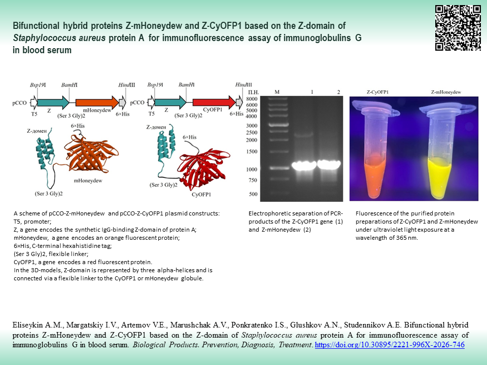
RESULTS. Genetic constructs pCCO-Z-mHoneydew and pCCO-Z-CyOFP1 under the control of the T5 promoter encode chimeric proteins with the following structure: Z-domain of protein A — flexible linker (Ser-Ser-Ser-Gly-Ser-Ser-Ser-Gly) — fluorescent protein mHoneydew or CyOFP1 — 6×His tag. Soluble proteins were obtained with a yield of ~37 mg/L (Z-mHoneydew) and ~6 mg/L (Z-CyOFP1) with a purity ≥90%. For the Z-mHoneydew protein, excitation and emission maxima were observed at 480 and 536 nm, respectively; a 26 nm blue shift in emission was observed relative to native mHoneydew (562 nm). For Z-CyOFP1, the excitation maximum was recorded at 515 nm; an 18 nm red shift in excitation was observed relative to native CyOFP1 (497 nm), while maintaining the emission maximum. Dose-dependent binding of IgG to the hybrid proteins was demonstrated using ELISA. A linear dependence was observed for Z-mHoneydew in the concentration range of 30–235 ng/μL (R²=0.96) and 15–250 ng/μL (R²=0.97) for Z-CyOFP1, confirming the capacity of the hybrid proteins to bind IgG. Immunofluorescence assay showed the feasibility of detecting IgG, specific to conjugate benzo[a]pyrene-bovine serum albumin, in rabbit serum using hybrid proteins. A linear relationship was observed for Z-mHoneydew in the serum dilution range of 1:10–1:160 (R²=0.92) and 1:10–1:80 (R²=0.98) for Z-CyOFP1, evidencing the suitability of hybrid proteins for detection of specific antibodies in blood serum.
CONCLUSIONS. Hybrid proteins Z-mHoneydew and Z-CyOFP1 retain fluorescent properties and the capacity to bind to IgG, including serum IgG, which is promising from the point of view of developing prototypes of universal immunofluorescent test systems.
Keywords
immunoglobulin G,
immunofluorescence,
immunoassay,
ELISA,
Z-domain,
protein A,
Staphylococcus aureus,
mHoneydew,
CyOFP1,
hybrid proteins
Funding: This study was conducted by the Federal State Scientific Institute, Federal Research Centre for Coal and Coal Chemistry, Siberian Branch of the Russian Academy of Sciences, Institute of Human Ecology as part of the research and development work under State Assignment No. 1023032300035-4-3.1.3.
About the Authors
A. M. Eliseykin
Federal State Scientific Institute, Federal Research Centre for Coal and Coal Chemistry, Siberian Branch of the Russian Academy of Sciences, Institute of Human Ecology
Russian Federation
Russian Federation
Alexey M. Eliseykin
10 Leningradsky Ave., Kemerovo 650065
I. V. Margatskiy
Federal State Scientific Institute, Federal Research Centre for Coal and Coal Chemistry, Siberian Branch of the Russian Academy of Sciences, Institute of Human Ecology
Russian Federation
Russian Federation
Ivan V. Margatskiy
10 Leningradsky Ave., Kemerovo 650065
V. E. Artemov
Federal State Scientific Institute, Federal Research Centre for Coal and Coal Chemistry, Siberian Branch of the Russian Academy of Sciences, Institute of Human Ecology
Russian Federation
Russian Federation
Vladislav E. Artemov
10 Leningradsky Ave., Kemerovo 650065
A. V. Marushchak
Federal State Scientific Institute, Federal Research Centre for Coal and Coal Chemistry, Siberian Branch of the Russian Academy of Sciences, Institute of Human Ecology
Russian Federation
Russian Federation
Anna V. Marushchak
10 Leningradsky Ave., Kemerovo 650065
I. S. Ponkratenko
Federal State Scientific Institute, Federal Research Centre for Coal and Coal Chemistry, Siberian Branch of the Russian Academy of Sciences, Institute of Human Ecology
Russian Federation
Russian Federation
Igor S. Ponkratenko
10 Leningradsky Ave., Kemerovo 650065
A. N. Glushkov
Federal State Scientific Institute, Federal Research Centre for Coal and Coal Chemistry, Siberian Branch of the Russian Academy of Sciences, Institute of Human Ecology
Russian Federation
Russian Federation
Andrey N. Glushkov, Dr. Sci. (Med.)
10 Leningradsky Ave., Kemerovo 650065
A. E. Studennikov
Federal State Scientific Institute, Federal Research Centre for Coal and Coal Chemistry, Siberian Branch of the Russian Academy of Sciences, Institute of Human Ecology
Russian Federation
Russian Federation
Artem E. Studennikov, Cand. Sci. (Biol.)
10 Leningradsky Ave., Kemerovo 650065
References
1. Ghagane S, Puranik S, Gan S, et al. Frontiers of monoclonal antibodies: Applications in medical practices. Hum Antibodies. 2017;26(3):135–42. https://doi.org/10.3233/HAB-170331
2. Sidorin E, Solov’eva T. IgG-binding proteins of bacteria. Biochemistry (Mosc). 2011;76(3):295–308. https://doi.org/10.1134/s0006297911030023
3. Cruz A, Boer M, Strasser J, et al. Staphylococcal protein A inhibits complement activation by interfering with IgG hexamer formation. Proc Natl Acad Sci USA. 2021;118(7):e2016772118. https://doi.org/10.1073/pnas.2016772118
4. Goding J. Use of staphylococcal protein A as an immunological reagent. J Immunol Methods. 1978;(20):241–53. https://doi.org/10.1016/0022-1759(78)90259-4
5. Bao L, Yang A, Liu Z, et al. Development of a mammalian cell-based ZZ display system for IgG quantification. BMC Biotechnol. 2023;23(1):1–10. https://doi.org/10.1186/s12896-023-00798-2
6. Devdariani ZL, Tereshkina NE, Taranenko TM, et al. Results of modeling experiments in designing immune-enzyme test-system for the detection of antibodies to Yersinia pestis F1 (ELISA-Ab-F1 Yersinia pestis). Problems of Particularly Dangerous Infections. 2013;(1):74–7 (In Russ.). https://doi.org/10.21055/0370-1069-2013-1-74-77
7. Mazigi O, Schofield P, Langley D, Christ D. Protein A superantigen: structure, engineering and molecular basis of antibody recognition. Protein Eng Des Sel. 2019;32(8):359–66. https://doi.org/10.1093/protein/gzz026
8. Braisted A, Wells J. Minimizing a binding domain from protein A. Proc Nat Acad Sci USA. 1996;93(12):5688–92. https://doi.org/10.1073/pnas.93.12.5688
9. Tashiro M, Tejero R, Zimmerman D, et al. High-resolution solution NMR structure of the Z domain of staphylococcal protein A. J Mol Biol. 1997;272(4):573–90. https://doi.org/10.1006/jmbi.1997.1265
10. Le Brun A, Shah D, Athey D, et al. Self-assembly of protein monolayers engineered for improved monoclonal immunoglobulin G binding. Int J Mol Sci. 2011;12(8):5157–67. https://doi.org/10.3390/ijms12085157
11. Viviani V, Silva J, Ho P. A novel brighter bioluminescent fusion protein based on ZZ domain and Amydetes vivianii firefly luciferase for immunoassays. Front Bioeng Biotechnol. 2021;9:755045. https://doi.org/10.3389/fbioe.2021.755045
12. Park J, Kim M, Jose J, Park M. Covalently immobilized regenerable immunoaffinity layer with orientation-controlled antibodies based on Z-domain autodisplay. Int J Mol Sci. 2021;23(1):459. https://doi.org/10.3390/ijms23010459
13. Jeon D, Pyun J, Jose J, Park M. A regenerative immunoaffinity layer based on the outer membrane of Z-domains autodisplaying E. coli for immunoassays and immunosensors. Sensors (Basel). 2018;18(11):4030. https://doi.org/10.3390/s18114030
14. Hajdu T, Rebenku I, Serrano Cano T, et al. Fluorescence labeling-induced structural rearrangement of a monoclonal IgG revealed by biophysical experiments and simulations. Int J Biol Macromol. 2025;321(Pt 2):146209. https://doi.org/10.1016/j.ijbiomac.2025.146209
15. Witting E, Hober S, Kanje S. Affinity-based methods for site-specific conjugation of antibodies. Bioconjug Chem. 2021;32(8):1515–24. https://doi.org/10.1021/acs.bioconjchem.1c00313
16. Ji P, Wang K, Zhang L, et al. A new nanobody-enzyme fusion protein-linked immunoassay for detecting antibodies against influenza A virus in different species. J Biol Chem. 2022;298(12):102709. https://doi.org/10.1016/j.jbc.2022.102709
17. Yu K, Liu C, Kim B, Lee D. Synthetic fusion protein design and applications. Biotechnol Adv. 2015;33(1):155–64. https://doi.org/10.1016/j.biotechadv.2014.11.005
18. Nienhaus K, Nienhaus G. Genetically encodable fluorescent protein markers in advanced optical imaging. Methods Appl Fluoresc. 2022;10(4):042002. https://doi.org/10.1088/2050-6120/ac7d3f
19. Liu M, Wang B, Wang F, et al. Soluble expression of single-chain variable fragment (scFv) in Escherichia coli using superfolder green fluorescent protein as fusion partner. Appl Microbiol Biotechnol. 2019;103(15):6071–9. https://doi.org/10.1007/s00253-019-09925-6
20. Lu Q, Li X, Zhao J, et al. Nanobody-horseradish peroxidase and -EGFP fusions as reagents to detect porcine parvovirus in the immunoassays. J Nanobiotechnology. 2020;18(1):7. https://doi.org/10.1186/s12951-019-0568-x
21. Ilgen P, Grotjohann T, Jans DC, et al. RESOLFT nanoscopy of fixed cells using a Z-domain based fusion protein for labelling. PLoS One. 2015;10(9):e0136233. https://doi.org/10.1371/journal.pone.0136233
22. Yu XT, Fu XY, Gao XY, et al. Fc-specific and covalent conjugation of a fluorescent protein to a native antibody through a photoconjugation strategy for fabrication of a novel photostable fluorescent antibody. Anal Bioanal Chem. 2021;413(3):945–53. https://doi.org/10.1007/s00216-020-03051-3
23. Glushkov AN, Kostyanko MV, Anosova TP, et al. The model for research of immunological images of chemical carcinogens. Russian Journal of Immunology. 2007;1(3–4):246–50 (In Russ.). EDN: SWMMFP
24. Kostyanko MV, Glushkov AN. Method of conjugate hapten-protein preparing. Patent of the Russian Federation No. 2141114; 1998 (In Russ.). EDN: JPYAJM
25. Shaner N, Campbell R, Steinbach P, et al. Improved monomeric red, orange and yellow fluorescent proteins derived from Discosoma sp. red fluorescent protein. Nat Biotechnol. 2004;22(12):1567–72. https://doi.org/10.1038/nbt1037
26. Chu J, Oh Y, Sens A, et al. A bright cyan-excitable orange fluorescent protein facilitates dual-emission microscopy and enhances bioluminescence imaging in vivo. Nat Biotechnol. 2016;34(7):760–7. https://doi.org/10.1038/nbt.3550
27. Gintsburg AL, Karjagina-Zhulina AS, Kormilitsyna MI, et al. Recombinant plasmid DNA encoding TUL4spCBD recombinant protein synthesis, m15[pREP4, pTUL4spCBD] Escherichia coli strain as producer of tul4spcbd recombinant protein, TUL4spCBD recombinant protein, method for production thereof, and method for production of specific antibody against TUL4spCBD protein. Patent of the Russian Federation No. 2270249; 2006 (In Russ.). EDN: VETEJD
28. Nandy S, Crum M, Wasden K, et al. Protein A-Nanoluciferase fusion protein for generalized, sensitive detection of immunoglobulin G. Anal Biochem. 2023;660:114929. https://doi.org/10.1016/j.ab.2022.114929
29. Huang QL, Chen C, Chen YZ, et al. Application to immunoassays of the fusion protein between protein ZZ and enhanced green fluorescent protein. J Immunol Methods. 2006;309(1–2):130–8. https://doi.org/10.1016/j.jim.2005.11.009
30. Chu J, Oh Y, Sens A, et al. A bright cyan-excitable orange fluorescent protein facilitates dual-emission microscopy and enhances bioluminescence imaging in vivo. Nat Biotechnol. 2016;34(7):760–7. https://doi.org/10.1038/nbt.3550
31. Bajar BT, Wang ES, Zhang S, et al. A guide to fluorescent protein FRET pairs. Sensors (Basel). 2016;16(9):1488. https://doi.org/10.3390/s16091488
Review
For citations:
Eliseykin A.M., Margatskiy I.V., Artemov V.E., Marushchak A.V., Ponkratenko I.S., Glushkov A.N., Studennikov A.E. Bifunctional hybrid proteins Z-mHoneydew and Z-CyOFP1 based on the Z-domain of Staphylococcus aureus protein A for immunofluorescence assay of immunoglobulins G in blood serum. Biological Products. Prevention, Diagnosis, Treatment. 2026;26(2):196-207. (In Russ.) https://doi.org/10.30895/2221-996X-2026-746
Views:
774
JATS XML





 Email this article