


Therapeutic glycoproteins: sample preparation for N-glycan profiling of monoclonal antibodies

https://doi.org/10.30895/2221-996X-2026-727

Abstract
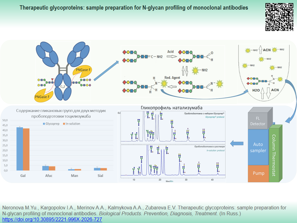
INTRODUCTION. Glycan profile is a critical quality parameter for therapeutic monoclonal antibodies (mAbs) that is consistently estimated during development and release of each drug batch. The need to develop a reproducible sample preparation protocol for glycan profiling brings about the relevance of the study. The profile should be adapted to the conditions of a standard physicochemical laboratory, so that it avoids using commercial preparation kits that are currently in short supply.
AIM. This study aimed to develop an alternative sample preparation procedure for quantitation of glycans in therapeutic monoclonal antibodies without using commercial kits.
MATERIALS AND METHODS. Omalizumab, ustekinumab, canakinumab, tocilizumab, natalizumab, and human anti-PD IgG2 were used as the study objects. N-glycans were released from mAbs by peptide-N-glycosidase (PNGase F) and labeled with fluorescent tags 2-aminobenzamide (2-АВ) or 2-aminobenzoic acid (2-АА) or InstantAB. Subsequently, glycan samples were cleaned from impurities. Glycan compounds were analyzed using hydrophilic interaction liquid chromatography (HILIC-FLD) on Alliance e2695 and Acquity Arc Bio chromatographic systems equipped with FLR 2475 fluorescence detector. The glycan profile was assessed by the content of functional glycan groups.
RESULTS. Conditions for preparing therapeutic mAbs N-glycans were chosen as follows: incubation with 2 mEU PNGase F per 100 µg protein in 10 mM Tris-HCl (pH 8.0) at 37 °C for 3 h without protein denaturation; derivatization of glycans with 2-AA at 65 °C for 1.5 h; extraction of 2-AA excess with acetonitrile. Centrifugation of labeled glycans with acetonitrile is suitable for purification and concentration of samples. Minor glycans with a content not more than 0.1–0.2% were determined using HILIC-FLD. Comparison with data obtained using a commercial kit for sample preparation indicated acceptable comparability of the results. When preparing the samples, we took into account structural features of specific mAbs, for instance, desialylation control during the staining was found necessary for high-sialylated mAbs.
CONCLUSIONS. The developed sample preparation procedure is suitable for the analysis of N-glycans of omalizumab, canakinumab and natalizumab and may be used to develop the analysis methods of the glycan profile of other glycoproteins using HILIC-FLD mode.
Keywords
monoclonal antibodies,
quality control,
HILIC,
2-aminobenzamide,
2-aminobenzoic acid,
liquid
chromatography,
sample preparation,
N-glycans,
glycoproteins
Funding: The study was conducted as part of applied research project funded by GENERIUM JSC.
About the Authors
M. Yu. Neronova
GENERIUM JSC
Russian Federation
Russian Federation
Maria Yu. Neronova
273 Zavodskaya St., Volginsky, Vladimir Region, 601125
I. A. Kargopolov
GENERIUM JSC
Russian Federation
Russian Federation
Ivan A. Kargopolov
273 Zavodskaya St., Volginsky, Vladimir Region, 601125
A. A. Merinov
GENERIUM JSC
Russian Federation
Russian Federation
Artem A. Merinov
273 Zavodskaya St., Volginsky, Vladimir Region, 601125
A. A. Kalmykova
GENERIUM JSC
Russian Federation
Russian Federation
Alena A. Kalmykova
273 Zavodskaya St., Volginsky, Vladimir Region, 601125
E. V. Zubareva
GENERIUM JSC
Russian Federation
Russian Federation
Ekaterina V. Zubareva
273 Zavodskaya St., Volginsky, Vladimir Region, 601125
References
1. Varki A, Cummings RD, Esko JD, et al. Essentials of glycobiology. 4th ed. Cold Spring Harbor (NY): Cold Spring Harbor Laboratory Press; 2022.
2. Mimura Y, Katoh T, Saldova R, et al. Glycosylation engineering of therapeutic IgG antibodies: Challenges for the safety, functionality and efficacy. Protein Cell. 2018;9(1):47–62. https://doi.org/10.1007/s13238-017-0433-3
3. Beck A, Debaene F, Diemer H, et al. Cutting-edge mass spectrometry characterization of originator, biosimilar and biobetter antibodies. J Mass Spectrom. 2015;50(2):285–97. https://doi.org/10.1002/jms.3554
4. Chirino AJ, Mire-Sluis A. Characterizing biological products and assessing comparability following manufacturing changes. Nat Biotechnol. 2004;22(11):1383–91. https://doi.org/10.1038/nbt1030
5. Zhou Q, Qiu H. The mechanistic impact of N-glycosylation on stability, pharmacokinetics, and immunogenicity of therapeutic proteins. J Pharm Sci. 2019;108(4):1366–77. https://doi.org/10.1016/j.xphs.2018.11.029
6. Majewska N, Tejada ML, Betenbaugh MJ, Agarwal N. N-Glyco sylation of IgG and IgG-like recombinant therapeutic proteins: Why is it important and how can we control it? Annu Rev Chem Biomol Eng. 2020;11:311–38. https://doi.org/10.1146/annurev-chembioeng-102419-010001
7. Hajba L, Szekrényes Á, Borza B, Guttman A. On the glycosylation aspects of biosimilarity. Drug Discov Today. 2018;23(3):616–25. https://doi.org/10.1016/j.drudis.2018.01.009
8. Houdebine LM. Production of pharmaceutical proteins by transgenic animals. Comp Immunol Microbiol Infect Dis. 2009;32(2):107–21. https://doi.org/10.1016/j.cimid.2007.11.005
9. Boune S, Hu P, Epstein AL, Khawli LA. Principles of N-linked glycosylation variations of IgG-based therapeutics: Pharmacokinetic and functional considerations. Antibodies (Basel). 2020;9(2):22. https://doi.org/10.3390/antib9020022
10. Ikegami T. Hydrophilic interaction chromatography for the analysis of biopharmaceutical drugs and therapeutic peptides: A review based on the separation characteristics of the hydrophilic interaction chromatography phases. J Sep Sci. 2019;42(1):130–213. https://doi.org/10.1002/jssc.201801074
11. De Leoz MLA, Duewer DL, Fung A, et al. NIST interlaboratory study on glycosylation analysis of monoclonal antibodies: Comparison of results from diverse analytical methods. Mol Cell Proteomics. 2020;19(1):11–30. https://doi.org/10.1074/mcp.RA119.001677
12. Cajic S, Hennig R, Grote V, et al. Removable dyes — the missing link for in-depth N-glycan analysis via multi-method approaches. Engineering. 2023;26(4):132–50. https://doi.org/10.1016/j.eng.2023.02.016
13. Kinoshita M, Yamada K. Recent advances and trends in sample preparation and chemical modification for glycan analysis. J Pharm Biomed Anal. 2021;207:114424. https://doi.org/10.1016/j.jpba.2021.114424
14. Keser T, Paviс T, Lauc G, Gornik O. Comparison of 2-aminobenzamide, procainamide and rapifluor-MS as derivatizing agents for high-throughput HILIC-UPLC-FLR-MS N-glycan analysis. Front Chem. 2018;6:324. https://doi.org/10.3389/fchem.2018.00324
15. Kameyama A, Dissanayake SK, Thet Tin WW. Rapid chemical de-N-glycosylation and derivatization for liquid chromatography of immunoglobulin N-linked glycans. PLoS One. 2018;13(5):e0196800. https://doi.org/10.1371/journal.pone.0196800
16. Zhang Q, Lasanajak Y, Song X. Oxidative release of natural glycans: Unraveling the mechanism for rapid N-glycan glycomics analysis. Anal Chem. 2024;96(42):16750–7. https://doi.org/10.1021/acs.analchem.4c03246
17. Magnelli P, Guthrie E, Taron CH, et al. Deglycosylation reagents and methods. US Patent No. US 9964548 B2; 2018. https://patents.google.com/patent/US9964548B2/en
18. Tiwold EK, Gyorgypal A, Chundawat SPS. Recent advances in biologic therapeutic N-glycan preparation techniques and analytical methods for facilitating biomanufacturing automation. J Pharm Sci. 2023;112(6):1485–91. https://doi.org/10.1016/j.xphs.2023.01.012
19. Anumula KR, Dhume ST. High resolution and high sensitivity methods for oligosaccharide mapping and characterization by normal phase high performance liquid chromatography following derivatization with highly fluorescent anthranilic acid. Glycobiology. 1998;8(7):685–94. https://doi.org/10.1093/glycob/8.7.685
20. Bigge JC, Patel TP, Bruce JA, et al. Nonselective and efficient fluorescent labeling of glycans using 2-amino benzamide and anthranilic acid. Anal Biochem. 1995;230(2):229–38. https://doi.org/10.1006/abio.1995.1468
21. Jeong YR, Kim SY, Park YS, Lee GM. Simple and robust N-glycan analysis based on improved 2-aminobenzoic acid labeling for recombinant therapeutic glycoproteins. J Pharm Sci. 2018;107(7):1831–41. https://doi.org/10.1016/j.xphs.2018.03.013
22. Kimzey M, Gyenes A. Compounds for labeling amine-containing compounds, and their use. US Patent No. US 20180156809 A1; 2018. https://patents.google.com/patent/US20180156809A1/en
23. Brousmiche DW, Yu Y-Q, Lauber M. Rapid fluorescence tagging of glycans and other biomolecules with enhanced MS signals. US Patent No. US 10436790 B2; 2014. https://patents.google.com/patent/US10436790
24. Baginski T. Compounds and methods for rapid labeling of N-glycans. US Patent No. US 8445292 B2; 2013. https://patents.google.com/patent/US8445292B2/en
25. Kimzey M. Use of mild electrophiles to reduce artifacts in analyzing glycans released from glycoproteins or glycopeptides. US Patent No. US 20190170763 A1; 2019. https://patents.google.com/patent/US20190170763A1/en
26. Stöckmann H, Duke RM, Millán Martín S, Rudd PM. Ultrahigh throughput, ultrafiltration-based n-glycomics platform for ultraperformance liquid chromatography (ULTRA(3)). Anal Chem. 2015;87(16):8316–22. https://doi.org/10.1021/acs.analchem.5b01463
27. Kinoshita M, Nakatani Y, Yamada K, et al. A rapid and facile preparation of APTS-labeled Nglycans by combination of ion pair-assisted extraction and HILIC-SPE for routine glycan analysis. J Pharm Biomed Anal. 2021;195:113875. https://doi.org/10.1016/j.jpba.2020.113875
28. Pabst M, Kolarich D, Pöltl G, et al. Comparison of fluorescent labels for oligosaccharides and introduction of a new postlabeling purification method. Anal Biochem. 2009;384(2):263–73. https://doi.org/10.1016/j.ab.2008.09.041
29. Xie Y, Mota LM, Bergin A, et al. High-throughput and high-sensitivity N-glycan profiling: A platform for biopharmaceutical development and disease biomarker discovery. Anal Biochem. 2021;623:114205. https://doi.org/10.1016/j.ab.2021.114205
30. Trbojević-Akmačić I, Lageveen-Kammeijer GSM, Heijs B, et al. High-throughput glycomic methods. Chem Rev. 2022;122(20):15865–913. https://doi.org/10.1021/acs.chemrev.1c01031
31. Sun X, Tao L, Yi L, et al. N-glycans released from glycoproteins using a commercial kit and comprehensively analyzed with a hypothetical database. J Pharm Anal. 2017;7(2):87–94. https://doi.org/10.1016/j.jpha.2017.01.004
32. Helali Y, Delporte C. Updates of the current strategies of labeling for N-glycan analysis. J Chromatogr B Analyt Technol Biomed Life Sci. 2024;1237:124068. https://doi.org/10.1016/j.jchromb.2024.124068
33. Aich U, Lakbub J, Liu A. State-of-the-art technologies for rapid and high-throughput sample preparation and analysis of N-glycans from antibodies. Electrophoresis. 2016;37(11):1468–88. https://doi.org/10.1002/elps.201500551
34. Reusch D, Haberger M, Maier B, et al. Comparison of methods for the analysis of therapeutic immunoglobulin G Fc-glycosylation profiles — part 1: Separation-based methods. MAbs. 2015;7(1):167–79. https://doi.org/10.4161/19420862.2014.986000
35. Segu Z, Stone T, Berdugo C, et al. A rapid method for relative quantification of N-glycans from a therapeutic monoclonal antibody during trastuzumab biosimilar development. mAbs. 2020;12(1):1750794. https://doi.org/10.1080/19420862.2020.1750794
36. Subedi GP, Barb AW. The structural role of antibody N-glycosylation in receptor interactions. Structure. 2015;23(9):1573–83. https://doi.org/10.1016/j.str.2015.06.015
37. Cantin G, Liu Q, Shah B, et al. Analytical and functional similarity of the biosimilar candidate ABP 654 to ustekinumab reference product. Drugs R D. 2023;23(4):421–38. https://doi.org/10.1007/s40268-023-00441-7
38. Alt N, Zhang TY, Motchnik P, et al. Determination of critical quality attributes for monoclonal antibodies using quality by design principles. Biologicals. 2016;44(5):291–305. https://doi.org/10.1016/j.biologicals.2016.06.005
39. Watanabe T, Inoue N, Kutsukake T, et al. Labeling conditions using a 2-aminobenzamide reagent for quantitative analysis of sialo-oligosaccharides. Biol Pharm Bull. 2000;23(3):269–73. https://doi.org/10.1248/bpb.23.269
40. Li M, Shen S, Letarte S, Flick T. Discovery and characterization of novel G0 glycan isomers in an afucosylated thera peutic antibody using liquid chromatography — mass spectro metry. J Am Soc Mass Spectrom. 2025;36(5):1034–40. https://doi.org/10.1021/jasms.4c00474
41. Askretkov AD, Shatalov DO. Development of a methodology for determining the glycosylation profile in eculizumab and other monoclonal antibodies. In: Mardanly SG, Pomazanov VV, Kiseleva VA, eds. Prospects for the introduction of innovative technologies in medicine and pharmacy. Orekhovo-Zuevo; 2021. P. 30–6 (In Russ.). EDN: NCQEMC
42. Zhang Q, Azzam T, Lasanajak Y, et al. Oxidative release of natural glycans (ORNG): Preparation of truncated N-glycans for engineering glycoproteins. Chem Eur J. 2025;31:e202501020. https://doi.org/10.1002/chem.202501020
Review
For citations:
Neronova M.Yu., Kargopolov I.A., Merinov A.A., Kalmykova A.A., Zubareva E.V. Therapeutic glycoproteins: sample preparation for N-glycan profiling of monoclonal antibodies. Biological Products. Prevention, Diagnosis, Treatment. (In Russ.) https://doi.org/10.30895/2221-996X-2026-727
Views:
266
JATS XML





 Email this article